ISSN 1004-4140
CN 11-3017/P
| Citation: |
WANG S G, PU R W, LIU Y J, et al. Optimization of Dual-energy Spectral Lower-extremity Computed Tomography Venography Scanning Protocol: Phantom Study[J]. CT Theory and Applications, 2025, 34(1): 73-81. DOI: 10.15953/j.ctta.2024.117. (in Chinese).

|
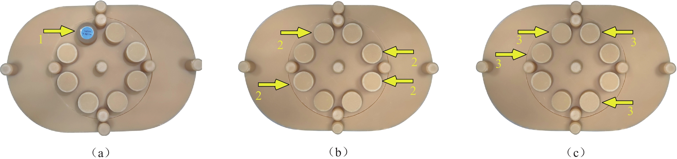
Objective: To optimize a scanning protocol for dual-energy spectral lower-extremity computed tomography venography (CTV) based on a phantom study. Methods: Test plugs were placed in the cavities of an energy CT quality-control phantom to simulate clinical scenarios. A 4 mgI/mL iodine rod was used to mimic venous enhancement in the lower extremities, and duck blood clots of various sizes were placed in test tubes containing 4 mgI/mL of iodine solution to simulate thrombi of different sizes in the lower-extremity veins. Revolution CT was used to perform standard CT imaging (Group A) and spectral imaging (Group B) on phantoms containing iodine rods and test tubes. The imaging parameters for Group A were as follows: tube voltage of 120 kVp, auto tube-current technology (100~600 mA), noise index (NI) of 10, and image reconstruction using 40% posterior multimodel adaptive statistical iterative reconstruction (ASIR-V). The imaging parameters for Group B were spectral imaging (GSI) mode, instantaneous dual tube voltage of 80/140 kVp, tube current with GSI Assist technology, and three scan groups based on NI values of 10, 11, and 12. For each scan group, monoenergetic images at 40~70 keV with 10 keV intervals were reconstructed, each combined with 40%, 60%, and 80% posterior ASIR-V, which resulted in 36 image sets. Other imaging parameters for Groups A and B were consistent. The effective radiation doses (ED) for Groups A and B were recorded after scanning, and the contrast-to-noise ratio (CNR) of the iodine rods was calculated. Subjective image quality and true- and false-positive rates for thrombus identification were assessed. Results: The EDs for Group B, with NI values of 11 and 12, were 21.5% and 32.2% lower than those for Group A, respectively. In Group B, for the scan groups with NI values of 10 and 11, except for the images at 70 keV combined with 40% ASIR-V and at 60 keV combined with 40% ASIR-V, the CNR of the iodine rods was higher than that in Group A. The highest edge-sharpness scores for the iodine rods in Group B were observed in the scan group with an NI value of 10 for images at 50 keV combined with 40% and 60% ASIR-V, and in the scan group with an NI value of 11 for images at 50 keV combined with 60% ASIR-V. These three image sets scored 5 (4, 5) compared with Group A’s score of 3 (3, 4). The true- and false-positive rates for large-thrombus identification in Group A were 65.0% and 30.0%, respectively, whereas those for small-thrombus identification were 55.0% and 50.0%, respectively. In Group B, the best thrombus-identification efficacy was observed in the scan groups, with NI values of 10 and 11 for images at 50 keV combined with 60% ASIR-V. The true- and false-positive rates for large-thrombus identification were 90.0% and 5.0%, respectively, whereas those for small-thrombus identification were 80.0% and 5.0%, respectively. Conclusions: Setting the NI to 11 and reconstructing monoenergetic images at 50 keV combined with 60% ASIR-V is the optimal imaging strategy for dual-energy spectral lower-extremity CTV, which balances between image quality and radiation dose in the current phantom study.
| [1] |
ROGNONI C, LUGLI M, MALETI O, et al. Clinical guidelines versus current clinical practice for the mana-gement of deep vein thrombosis[J]. Journal of Vascular Surgery: Venous and Lymphatic Disorders, 2021, 9(5): 1334-1344. DOI: 10.1016/j.jvsv.2021.01.020.
|
| [2] |
KAKKOS S K, GOHEL M, BAEKGAARD N, et al. Editor’s choice-european society for vascular surgery (ESVS) 2021 clinical practice guidelines on the management of venous thrombosis[J]. European Journal of Vascular and Endovascular Surgery, 2021, 61(1): 9-82. DOI: 10.1016/j.ejvs.2020.09.023.
|
| [3] |
中国医师协会血管外科医师分会静脉学组. 常见静脉疾病诊治规范(2022年版)[J]. 中华血管外科杂志, 2022, 7(1): 12-29. DOI: 10.3877/cma.j.issn.1674-0793.2022.04.002.
|
| [4] |
TANOUE S, NAKAURA T, IYAMA Y, et al. Diagnostic performance of dual-layer computed tomography for deep vein thrombosis in indirect computed tomography venography[J]. Circulation Journal, 2020, 84(4): 636-641. DOI: 10.1253/circj.CJ-19-0722.
|
| [5] |
王诗耕, 刘义军, 李贝贝, 等. 下肢能谱CT静脉成像最佳重建能级和自适应统计迭代重建权重[J]. 中国介入影像与治疗学, 2023, 20(10): 625-629. DOI: 10.13929/j.issn.1672-8475.2023.10.012.
WANG S G, LIU Y J, LI B B, et al. Optimal reconstructive combination of keV and adaptive statistical iterative reconstruction-V level for energy spectral CT venography of lower extremity[J]. Chinese Journal of Interventional Imaging and Therapy, 2023, 20(10): 625-629. DOI: 10.13929/j.issn.1672-8475.2023.10.012. (in Chinese).
|
| [6] |
王伟新, 张秋奂, 郭鹏德, 等. 能谱CT单能量结合ASiR技术对腹部静脉成像质量的优化研究[J]. CT理论与应用研究, 2019, 28(1): 61-72. DOI: 10.15953/j.1004-4140.2019.28.01.07.
WANG W X, ZHANG Q H, GUO P D, et al. The evaluation of imaging quality of abdominal vein by combining spectral CT optimal monotromatic imging and ASiR technique[J]. CT Theory and Applications, 2019, 28(1): 61-72. DOI: 10.15953/j.1004-4140.2019.28.01.07. (in Chinese).
|
| [7] |
王宏, 李玲, 逯瑶, 等. 乳腺癌颈胸腹盆增强CT: 能谱与常规扫描模式比较[J]. CT理论与应用研究, 2022, 31(4): 489-498. DOI: 10.15953/j.ctta.2022.094.
WANG H, LI L, LU Y, et al. Neck-chest-abdomen-pelvis combined enhanced CT in breast cancer patients: Comparison between dual-energy and conventional scanning mode[J]. CT Theory and Applications, 2022, 31(4): 489-498. DOI: 10.15953/j.ctta.2022.094. (in Chinese).
|
| [8] |
WOO H, CHAI J W, CHOI Y H, et al. Metal artifact reduction for orthopedic prosthesis in lower extremity CT venography: Evaluation of image quality and vessel conspicuity[J]. Cardiovascular and Interventional Radiology, 2019, 42(11): 1619-1626. DOI: 10.1007/s00270-019-02326-2.
|
| [9] |
ZHAO F L, GOU Y L, LAN Y S. Experimental study on the effects of different exposure conditions and contrast agent concentrations on spectral computed tomography virtual non-contrast images[J]. Quantitative Imaging in Medicine and Surgery, 2024, 14(1): 986-994. DOI: 10.21037/qims-23-1092.
|
| [10] |
KIM K A, CHOI S Y, KIM R. Endovascular treatment for lower extremity deep vein thrombosis: An overview[J]. Korean Journal of Radiology, 2021, 22(6): 931-943. DOI: 10.3348/kjr.2020.0675.
|
| [11] |
王诗耕, 刘义军, 李贝贝, 等. 能谱CT单能级成像结合ASIR-V对下肢深静脉血栓清晰度的影响[J]. 临床放射学杂志, 2023, 42(8): 1337-1343. DOI: 10.13437/j.cnki.jcr.2023.08.020.
WANG S G, LIU Y J, LI B B, et al. The effect of virtual monochromatic images on the clarity of lower extremity deep vein thrombosis with spectral CTV combined with ASIR-V reconstruction strategy[J]. Journal of Clinical Radiology, 2023, 42(8): 1337-1343. DOI: 10.13437/j.cnki.jcr.2023.08.020. (in Chinese).
|
| [12] |
中华医学会放射学分会, 中国医师协会放射医师分会, 安徽省影像临床医学研究中心. 能量CT临床应用中国专家共识[J]. 中华放射学杂志, 2022, 56(5): 476-487. DOI: 10.3760/cma.j.cn112149-20220118-00051.
|
| [13] |
刘娜, 周宇婧, 赵明月, 等. 头颈部CTA中GSI Assist单能量成像结合个体化对比剂注射方案的应用研究[J]. 临床放射学杂志, 2022, 41(4): 741-746. DOI: 10.13437/j.cnki.jcr.2022.04.013.
LIU N, ZHOU Y J, ZHAO M Y, et al. Application study of the GSI monoenergetic imaging combined with individualized contrast injection scheme in head and neck CT angiography[J]. Journal of Clinical Radiology, 2022, 41(4): 741-746. DOI:10.13437/j.cnki.jcr.2022.04.013. (in Chinese).
|
| [14] |
GAUNTT D M. A suggested method for setting up GSI profiles on the GE Revolution CT scanner[J]. Journal of Applied Clinical Medical Physics, 2019, 20(12): 169-179. DOI: 10.1002/acm2.12754.
|
| [15] |
王永胜, 张鹏宇, 李方中, 等. 不同管电压对头颈部CTA图像质量影响的研究[J]. CT理论与应用研究, 2022, 31(5): 631-638. DOI: 10.15953/j.ctta.2022.026.
WANG Y S, ZHANG P Y, LI F Z, et al. Study on the influence of head and neck CTA image quality under different tube voltages[J]. CT Theory and Applications, 2022, 31(5): 631-638. DOI: 10.15953/j.ctta.2022.026. (in Chinese).
|
| [16] |
孙杰, 倪亚博, 沈云, 等. 联合全新迭代算法(ASiR-V)的Revolution CT低keV单能量优化腹部血管成像的价值[J]. 放射学实践, 2022, 37(10): 1238-1242. DOI: 10.13609/j.cnki.1000-0313.2022.10.009.
SUN J, NI Y B, SHEN Y, et al. The optimization abdominal angiography value of revolution CT low-keV single energy combines with the new iterative algorithm (ASIR-V)[J]. The American Journal of Cardio-logy, 2022, 37(10): 1238-1242. DOI: 10.13609/j.cnki.1000-0313.2022.10.009. (in Chinese).
|
| [1] | ZANG Yikai, LI Jing, LU Xiuliang, YAN Cheng. Image Quality Inprovement for Small Vessel in Diabetic Foot Arteriography Using Dual-energy Computed Tomography[J]. CT Theory and Applications, 2025, 34(1): 83-88. DOI: 10.15953/j.ctta.2024.130 |
| [2] | WANG Yongsheng, YANG Leiqing, YANG Yifan, PEI Qingxia, WANG Chensi, LU Mengyun, HE Junlin, CHEN Wenjing, TIAN Xiangbao. The Effect of Different Trigger Thresholds on the Quality of Pulmonary Artery CT Angiography Images[J]. CT Theory and Applications, 2024, 33(2): 175-181. DOI: 10.15953/j.ctta.2023.121 |
| [3] | FANG Shu, CHEN Linyu, CHEN Yong, DONG Haipeng, WANG Lan, MIN Jihua. Phantom Study on the Influence of CT Model and Tube Voltage on Image Quality[J]. CT Theory and Applications, 2022, 31(3): 345-350. DOI: 10.15953/j.ctta.2021.042 |
| [4] | YANG Zhengjun, ZHANG Ang, CHEN Yong, JIANG Jiang, WANG Lingyun, ZHANG Yong, ZHANG Xuan, QI Xiaofeng. The Effect of Radiation Dose and Tube Potential on Image Quality of CT: A Task-based Image Quality Assessment[J]. CT Theory and Applications, 2022, 31(2): 211-217. DOI: 10.15953/j.ctta.2021.060 |
| [5] | XIA Zhenying, WU Dan, YU Jianan, LI Siyuan, SONG Wenyan. The Influence of Different Noise Indexes on Chest CT Image Quality for Patients with PCP[J]. CT Theory and Applications, 2019, 28(2): 221-228. DOI: 10.15953/j.1004-4140.2019.28.02.08 |
| [6] | WANG Weixin, ZHANG Qiuhuan, GUO Pengde, HE Feifei, LIU Ming, LI Jie, CHEN Zhengguang. The Evaluation of Imaging Quality of Abdominal Vein by Combining Spectral CT Optimal Monotromatic Imging and ASiR Technique[J]. CT Theory and Applications, 2019, 28(1): 61-72. DOI: 10.15953/j.1004-4140.2019.28.01.07 |
| [7] | BU Yu-lian, ZHANG Huan, PAN Zi-lai, YANG Wen-jie, CHEN Ke-min, YAN Fu-hua. The Study of the Image Quality of Dual Energy Spectral CT in the Assessment of Myocardial Infarction[J]. CT Theory and Applications, 2016, 25(3): 279-286. DOI: 10.15953/j.1004-4140.2016.25.03.04 |
| [8] | SHAO Jun-ming, XU Xiao-dong, KONG Jun. Research on CT System Imaging Quality[J]. CT Theory and Applications, 2006, 15(3): 61-67. |
| [9] | LI Jian-ying. GEVCT–Harmony Between High Image Quality and Low X-ray Dose[J]. CT Theory and Applications, 2006, 15(3): 57-60. |
| [10] | LI Da-sheng, SUN Yong-guang. Development of Diagnosis in Lower Extremities Deep Venous Thrombosis by CT Venography[J]. CT Theory and Applications, 2004, 13(4): 40-44. |
Supported by: Beijing Renhe Information Technology Co. Ltd 


 DownLoad: