ISSN 1004-4140
CN 11-3017/P

| Citation: |
YAN W, JIN E H, WEI W, et al. Exploring the Influencing Factors of Acute Necrotic Accumulation Outcome[J]. CT Theory and Applications, xxxx, x(x): 1-6. DOI: 10.15953/j.ctta.2024.295. (in Chinese).

|
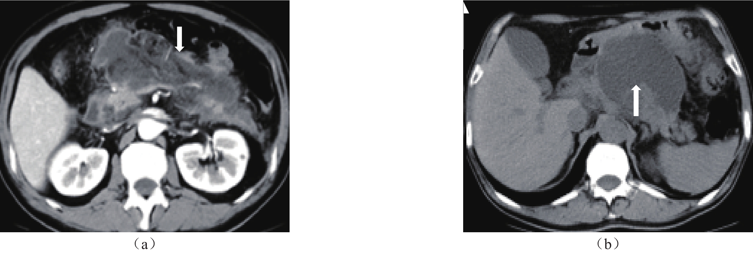
To explore the independent risk factors and predictive efficacy of ANC conversion to encapsulated necrosis (WON). Methods: A Retrospective analysis of CT/MRI features in 53 cases of ANC, divided into the WON and absorption groups, based on the outcome after 4 weeks of ANC formation. The chi square test or t-test were used to compare the statistical significance of differences in etiology and laboratory tests between the two groups. Regression analysis was used to identify independent risk factors affecting the outcome of ANC. Receiver operating characteristic (ROC) curves were used to obtain the area under the curve (AUC) and evaluate the predictive efficacy of each risk factor for the outcome of ANC to WON.
Logistic regression analysis showed that the P-values for necrotic volume ≥ 30% and MCTSI score > 6 were both < 0.05, with OR values of 9.21 and 16.04, respectively. ROC curve analysis showed that the P-values for necrotic volume ≥ 30% and MCTSI score> 6 were both < 0.05, with AUC values of 0.86 and 0.88, respectively.
Necrosis volume ≥ 30% and MCTSI score > 6 points are independent risk factors for the progression of ANC into WON, and the predictive performance of both is significant.
| [1] |
LANKISCH P G, APTE M, BANKS P A. Acute pancreatitis[J]. Lancet, 2015, 386(9988): 85-96. DOI: 10.1016/S0140-6736(14)60649-8.
|
| [2] |
PETROV M S, YADAV D. Global epidemiology and holistic prevention of pancreatitis[J]. Nature Reviews Gastroenterology & Hepatology, 2019, 16(3): 175-184. DOI: 10.1038/s41575-018-0087-5.
|
| [3] |
HABTEZION A, GUKOVSKAVA A S, PANDOL S J. Acute Pancreatitis: A Multifaceted Set of Organelle and Cellular Interactions[J]. Gastroenterology, 2019, 156(7): 1941-1950. DOI: 10.1053/j.gastro.2018.11.082.
|
| [4] |
BANKS P A, BOLLEN T L, DERVENIS C, et al. Classification of acute pancreatitis--2012: revision of the Atlanta classification and definitions by international consensus[J]. Gut, 2013, 62(1): 102-111. DOI: 10.1136/gutjnl-2012-302779.
|
| [5] |
GARDNER T B. Acute Pancreatitis[J]. Ann Intern Med, 2021, 174(2): C17-C32. DOI: 10.7326/AITC202102160.
|
| [6] |
HUANG J, QU H P, ZHENG Y F, et al. The revised Atlanta criteria 2012 altered the classification, severity assessment and management of acute pancreatitis[J]. Hepatobiliary Pancreat Diseases International, 2016, 15(3): 310-315. DOI: 10.1016/s1499-3872(15)60040-6.
|
| [7] |
闫威, 董力宁, 张斌斌, 等. 急性胰腺炎患者坏死性积聚的CT和MRI特征及转归分析[J]. CT理论与应用研究, 2023, 32(01): 113-120. DOI: 10.15953/j.ctta.2022.141.
YAN W, DONG L N, ZHANG B B, et al. CT and MRI features and outcome analysis of necrotic accumulation in patients with acute pancreatitis[J]. Research on CT Theory and Application, 2023, 32(01): 113-120. DOI: 10.15953/j.ctta.2022.141.
|
| [8] |
DIMAIO C J. Management of complications of acute pancreatitis[J]. Current Opinion Gastroenterol, 2018, 34(5): 336-342. DOI: 10.1097/MOG.0000000000000462.
|
| [9] |
GRASSEDONIO E, TOIA P, LA G L, et al. Role of computed tomography and magnetic resonance imaging in local complications of acute pancreatitis[J]. Gland Surgery, 2019, 8(2): 123-132. DOI: 10.21037/gs.2018.12.07.
|
| [10] |
TAYDAS O, UNAL E, KARAOSMANOGLU A D, et al. Accuracy of early CT findings for predicting disease course in patients with acute pancreatitis[J]. Japanese journal of radiology, 2018, 36(2): 151-158. DOI: 10.1007/s11604-017-0709-9.
|
| [11] |
RANA S S, SHARMA R K, GUPTA P, et al. Natural course of asymptomatic walled off pancreatic necrosis[J]. Digestive and Liver Disease, 2019, 51(5): 730-734. DOI: 10.1016/j.dld.2018.10.010.
|
| [12] |
闫媛媛, 靳二虎, 张洁, 等. CT和MRI对急性胰腺炎局部并发症的诊断价值研究[J]. CT理论与应用研究, 2018, 27(03): 393-400. DOI: 10.15953/j.1004-4140.2018.27.03.13.
YAN Y Y, JIN E H, ZHANG J, et al. Research on the diagnostic value of CT and MRI for local complications of acute pancreatitis[J]. Research on CT Theory and Application, 2018, 27(03): 393-400. DOI: 10.15953/j.1004-4140.2018.27.03.13.
|
| [13] |
THOENI R F. Imaging of Acute Pancreatitis[J]. Radiologic clinics of North America, 2015, 53(6): 1189-1208. DOI: 10.1016/j.rcl.2015.06.006.
|
| [14] |
BEZMAREVIC M, VANDIJK S M, VOERMANS R P, ETAL. Management of (Peri)Pancreatic Collections in Acute Pancreatitis[J]. Visceral Medicine, 2019, 35(2): 91-96. DOI: 10.1159/000499631.
|
| [15] |
MANRAI M, KOCHHAR R, GUPTA V, et al. Outcome of Acute Pancreatic and Peripancreatic Collections Occurring in Patients With Acute Pancreatitis[J]. Annals of Surgery, 2018, 267(2): 357-363. DOI: 10.1097/SLA.0000000000002065.
|
| [16] |
SARATHI P P, DAS K, BHATTACHARYYA A, et al. Natural resolution or intervention for fluid collections in acute severe pancreatitis[J]. British Journal of Surgery, 2014, 101(13): 1721-1728. DOI: 10.1002/bjs.9666.
|
| [17] |
刘建, 李昂, 刘殿刚, 等. CT检查预测急性胰腺炎局部并发症转归的价值[J]. 中华普外科手术学杂志(电子版), 2017, 11(04): 285-288.
LIU J, LI A, LIU D G, et al. The value of CT examination in predicting the outcome of local complications in acute pancreatitis[J]. Chinese Journal of General Surgery (Electronic Edition), 2017, 11(04): 285-288.
|
| [18] |
SUREKA B, BANSAL K, PATIDAR Y, et al. Imaging lexicon for acute pancreatitis: 2012 Atlanta Classification revisited[J]. Gastroenterology report, 2016, 4(1): 16-23. DOI: 10.1093/gastro/gov036.
|
| [19] |
MEDEROS M A, REBER H A, GIRGIS M D. Acute Pancreatitis: A Review[J]. Journal of the American Medical Association, 2021, 325(4): 382-390. DOI: 10.1001/jama.2020.20317.
|
| [20] |
ALBERTI P, PANDO E, MATA R, et al. Evaluation of the modified computed tomography severity index (MCTSI) and computed tomography severity index (CTSI) in predicting severity and clinical outcomes in acute pancreatitis[J]. Journal of Digestive Diseases, 2021, 22(1): 41-48. DOI: 10.1111/1751-2980.12961.
|
| [21] |
YAMAMIYA A, KITAMUTA K, YOSHIDA H, et al. Prediction of the progression of walled-off necrosis in patients with acute pancreatitis on whole pancreatic perfusion CT[J]. Journal of Hepatobiliary Pancreat Sciences, 2020, 27(10): 739-746. DOI: 10.1002/jhbp.803.
|
Supported by: Beijing Renhe Information Technology Co. Ltd 


 DownLoad: