ISSN 1004-4140
CN 11-3017/P

| Citation: |
QI R F, JIANG W L, HAO Y Y, et al. A Study of Image Quality and Radiation Dose in Lower Extremity Computed Tomography Angiography Using Caudo-cranial Flash Scanning[J]. CT Theory and Applications, 2025, 34(3): 1-7. DOI: 10.15953/j.ctta.2024.210. (in Chinese).

|
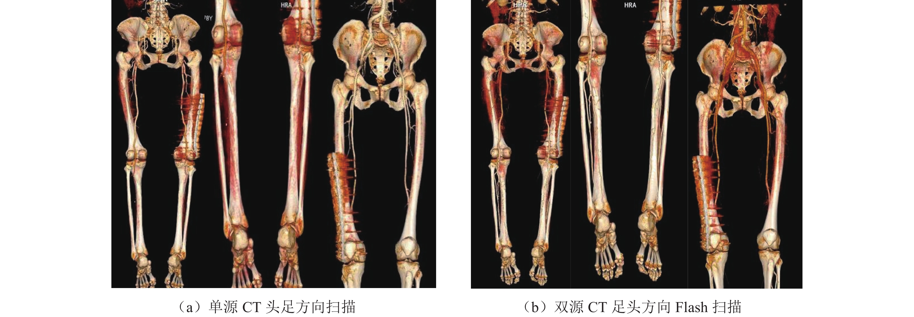
Objective: We conducted a comparative study of image quality and radiation dose in CTA angiography of lower extremity arteries with single-source CT cranio-cauda and dual-source CT cranio-cauda Flash scans. Methods: This prospective study enrolled 50 patients with suspected peripheral occlusive arterial disease which required CTA imaging of the lower extremities. Patients were randomly assigned to a control group (P1) or an experimental group (P2). Group P1 was scanned with protocol 1: single-source CT cranio-cauda direction, Group P2 was scanned with protocol 2: dual-source CT cranio-cauda direction Flash. Intravascular CT values, signal-to-noise ratio (SNR), and contrast-to-noise ratio (CNR) were compared as a group. Image quality was assessed by two radiologists. CT volume dose index (CTDIVOI) and dose length product (DLP) were compared as well. Results: Statistical differences were observed between P1 and P2 groups in intravascular CT values, SNR, and CNR at the aorta, external iliac artery, popliteal artery, and anterior tibial artery. No statistically significant difference was seen in intravascular CT values, SNR, or CNR at the femoral artery. The mean intravascular CT value in the P2 group was higher than that in the P1 group ((534.4±25.2) vs. (480.6±143.4)), and showed better consistency in each part. In the subjective evaluation, significant differences were found in image quality scores for the aorta and inferior knee arteries between P1 and P2 groups, but no significant differences between groups were found in image quality scores for the femoral artery and popliteal artery The radiation dose in the P2 group was significantly lower than that in the P1 group, including a 64.0% reduction in CTDIvol ((0.9±0.3) mGy to (0.5±0.3) mGy) and a 63.4% reduction in DLP ((113.5±33.4) mGy·cm to (310.1±53.5) mGy·cm). Conclusion: Lower extremity CTA combined with dual-source CT caudo-cranial Flash scanning yields better quality images in the small arterial system below the knee with higher CT values, SNR, and CNR, and better consistency than single-source CT cranio-cauda scanning. This method also reduces the radiation dose for lower extremity CTA examinations.
| [1] |
MCDERMOTT M M. The magnitude of the problem of peripheral arterial disease: Epidemiology and clinical significance[J]. Cleveland Clinic Journal of Medicine, 2006, 73(S4): S2-S7.
|
| [2] |
MARGOLIS J, BARRON J J, GROCHULSKI W D. Health care resources and costs for treating peripheral artery disease in a managed care population: Results from analysis of administrative claims data[J]. Journal of Managed Care Pharmacy, 2005, 11(9): 727-734. DOI: 10.18553/jmcp.2005.11.9.727.
|
| [3] |
HEIJENBROK-KAL M H, KOCK M C, HUNINK M G. Lower extremity arterial disease: Multidetector CT angiography meta-analysis[J]. Radiology, 2007, 245(2): 433-439. DOI: 10.1148/radiol.2451061280. (in Chinese).
|
| [4] |
中华医学会放射学分会, 下肢动脉CTA扫描技术专家共识协作组, 金征宇. 下肢动脉CT血管成像扫 描技术专家共识[J]. 中华放射学杂志, 2019, 53(2): 88-92. DOI: 10.3760/cma.j.issn.1005-1201.2019.02.002.
Chinese Society of Radiology C M A, Lower Limb Artery CTA Scanning Technology Expert Consensus Collaboration Group, JIN Z Y. Expert consensus of lower extremity CT angiography[J]. Chinese Journal of Radiology, 2019, 53(2): 88-92. DOI: 10.3760/cma.j.issn.1005-1201.2019.02.002. (in Chinese).
|
| [5] |
RUBIN G D, SCHMIDT A J, LOGAN L J, et al. Multi-detector row CT angiography of lower extremity arterial inflow and runoff: Initial experience[J]. Radiology, 2001, 221(1): 146-158. DOI: 10.1148/radiol.2211001325.
|
| [6] |
SIRIAPISITH T, WASINRAT J, MUTIRANGURA P, et al. Optimization of the table speed of lower extremity CT angiography protocols in different patient age groups[J]. Journal of Cardiovascular Computed Tomography, 2010, 4(3): 173-183. DOI: 10.1016/j.jcct.2010.03.011.
|
| [7] |
WINTERSPERGER B, JAKOBS T, HERZOG P, et al. Aorto-iliac multidetector-row CT angiography with low kV settings: Improved vessel enhancement and simultaneous reduction of radiation dose[J]. European Radiology, 2005, 15(2): 334-341. DOI: 10.1007/s00330-004-2575-y.
|
| [8] |
朱蕾, 牛延涛, 张永县, 等. 不同迭代重建算法在眼眶CT中的适用性研究[J]. CT理论与应用研究(中英文), 2024, 33(4): 487-496. DOI: 10.15953/j.ctta.2024.045.
ZHU L, NIU Y T, ZHANG Y X, et al. Applicability of different iterative reconstruction algorithms in orbital computed tomography[J]. CT Theory and Applications, 2024, 33(4): 487-496. DOI: 10.15953/j.ctta.2024.045. (in Chinese).
|
| [9] |
LIU B, GAO S, CHANG Z, et al. Lower extremity CT angiography at 80 kVp using iterative model reconstruction[J]. Diagnostic and Interventional Imaging, 2018, 99(9): 561-568. DOI: 10.1016/j.diii.2018.04.006.
|
| [10] |
OFER A, NITECKI S S, LINN S, et al. Multidetector CT angiography of peripheral vascular disease: A prospective comparison with intraarterial digital subtraction angiography[J]. American Journal of Roentgenology, 2003, 180(3): 719-724. DOI: 10.2214/ajr.180.3.1800719.
|
| [11] |
QANADLI S D, CHIAPPORI V, KELEKIS A. Multislice computed tomography of peripheral arterial disease: New approach to optimize vascular opacification with 16-row platform[J]. European Radiology, 2004, 14(S2): b806.
|
| [12] |
WILLMANN J K, MAYER D, BANYAI M, et al. Evaluation of peripheral arterial bypass grafts with multi-detector row CT angiography: Comparison with duplex US and digital subtraction angiography[J]. Radiology, 2003, 229(2): 465-474. DOI: 10.1148/radiol.2292021123.
|
| [13] |
BRUDER H, PETERSILKA M, MEHLDAU H, et al. Flash imaging in dual source CT (DSCT)[C]//SPIE Medical Imaging, Florida, United States, 2009: 138-148.
|
| [14] |
MEYER M, HAUBENREISSER H, SCHOEPF U J, et al. Closing in on the K edge: Coronary CT angiography at 100, 80, and 70 kV-initial comparison of a second- versus a third-generation dual-source CT system[J]. Radiology, 2014, 273(2): 373-382. DOI: 10.1148/radiol.14140244.
|
| [15] |
BUI T D, GELFAND D, WHIPPLE S, et al. Comparison of CT and catheter arteriography for evaluation of peripheral arterial disease[J]. Vascular and Endovascular Surgery, 2005, 39(6): 481-490. DOI: 10.1177/153857440503900604.
|
| [16] |
POLLAK A W, NORTON P T, KRAMER C M. Multimodality imaging of lower extremity peripheral arterial disease: current role and future directions[J]. Circulation Cardiovascular imaging, 2012, 5(6): 797-807. DOI: 10.1161/CIRCIMAGING.111.970814.
|
| [17] |
MET R, BIPAT S, LEGEMATE D A, et al. Diagnostic performance of computed tomography angiography in peripheral arterial disease: A systematic review and meta-analysis[J]. Journal of the American Medical Association, 2009, 301(4): 415-424. DOI: 10.1001/jama.301.4.415.
|
| [18] |
KOCK M C, ADRIAENSEN M E, PATTYNAMA P M, et al. DSA versus multi-detector row CT angiography in peripheral arterial disease: Randomized controlled trial[J]. Radiology, 2005, 237(2): 727-737. DOI: 10.1148/radiol.2372040616.
|
| [19] |
FLEISCHMANN D. CT angiography: Injection and acquisition technique[J]. Radiologic Clinics of North America, 2010, 48(2): 237-247. DOI: 10.1016/j.rcl.2010.02.002.
|
| [20] |
LESCHKA S, STOLZMANN P, SCHMID F T, et al. Low kilovoltage cardiac dual-source CT: Attenuation, noise, and radiation dose[J]. European Radiology, 2008, 18(9): 1809-1817. DOI: 10.1007/s00330-008-0966-1.
|
| [21] |
LAYRITZ C, MUSCHIOL G, FLOHR T, et al. Automated attenuation-based selection of tube voltage and tube current for coronary CT angiography: Reduction of radiation exposure versus a BMI-based strategy with an expert investigator[J]. Journal of Cardiovascular Computed Tomography, 2013, 7(5): 303-310. DOI: 10.1016/j.jcct.2013.08.010.
|
| [22] |
MOSCARIELLO A, TAKX R A, SCHOEPF U J, et al. Coronary CT angiography: Image quality, diagnostic accuracy, and potential for radiation dose reduction using a novel iterative image reconstruction technique-comparison with traditional filtered back projection[J]. European Radiology, 2011, 21(10): 2130-2138. DOI: 10.1007/s00330-011-2164-9.
|
| [23] |
OTA H, TAKASE K, RIKIMARU H, et al. Quantitative vascular measurements in arterial occlusive disease[J]. Radiographics: A Review Publication of the Radiological Society of North America, Inc, 2005, 25(5): 1141-1158. DOI: 10.1148/rg.255055014.
|
| [24] |
de ZORDO T, Von LUTTEROTTI K, DEJACO C, et al. Comparison of image quality and radiation dose of different pulmonary CTA protocols on a 128-slice CT: High-pitch dual source CT, dual energy CT and conventional spiral CT[J]. European Radiology, 2012, 22(2): 279-286. DOI: 10.1007/s00330-011-2251-y.
|
| [25] |
QI L, MEINEL F G, ZHOU C S, et al. Image quality and radiation dose of lower extremity CT angiography using 70 kVp, high pitch acquisition and sinogram-affirmed iterative reconstruction[J]. PloS One, 2014, 9(6): e99112. DOI: 10.1371/journal.pone.0099112.
|
| [26] |
QI L, ZHAO Y, ZHOU C S, et al. Image quality and radiation dose of lower extremity CT angiography at 70 kVp on an integrated circuit detector dual-source computed tomography[J]. Acta Radiologica, 2015, 56(6): 659-665. DOI: 10.1177/0284185114535391.
|
| [1] | QI Weiwei, CHENG Jin, CHEN Chuhan, AN Bei, LIU Xiaoyi, FU Ling, WANG Yi. Value of Low Tube Voltage Combined with Deep Learning Image Reconstruction Algorithm to Reduce Radiation Dose in Combined Thoracoabdominal Enhanced CT[J]. CT Theory and Applications. DOI: 10.15953/j.ctta.2025.001 |
| [2] | Peng Lexin¹, Ma Zixuan², Niu Yantao², Zhang Yongxian², Hu Lingjing¹, Wu Ning¹, Gao Xing³, Liu Dandan. The Effect of X-ray Energy on Dosimeter Measurements of CT Radiation Dose[J]. CT Theory and Applications. DOI: 10.15953/j.ctta.2024.315 |
| [3] | NI Xiaolong, ZHOU Fengyun, SHI Zheng, MA Yongzhong, CHENG Xiaoguang. Study on the Radiation Dose Distribution of Abdominal CT Scans Based on Dose Simulation Humans[J]. CT Theory and Applications, 2024, 33(6): 808-814. DOI: 10.15953/j.ctta.2024.087 |
| [4] | YANG Zhengjun, ZHANG Ang, CHEN Yong, JIANG Jiang, WANG Lingyun, ZHANG Yong, ZHANG Xuan, QI Xiaofeng. The Effect of Radiation Dose and Tube Potential on Image Quality of CT: A Task-based Image Quality Assessment[J]. CT Theory and Applications, 2022, 31(2): 211-217. DOI: 10.15953/j.ctta.2021.060 |
| [5] | WANG Wenjie, CHEN Ping, PAN Jinxiao, LI Yihong. Dual-energy CT Imaging Method Based on Reference Components[J]. CT Theory and Applications, 2021, 30(1): 61-69. DOI: 10.15953/j.1004-4140.2021.30.01.06 |
| [6] | LIU Wangyang, SUN Cunjie, ZHAO Honglan, WANG Xiuling. Study on Evaluation Method of Radiation Dose of Patients in TACE of IVR-CT[J]. CT Theory and Applications, 2019, 28(6): 653-658. DOI: 10.15953/j.1004-4140.2019.28.06.02 |
| [7] | LIANG Hong-sheng, CHEN Jin-jun, CHEN Xiao-yu, LIANG Yu-hao, OU Jian-yang, MA Guan-hua. Dose 4D CARE Technology Combined with Low kV and Low Dose Contrast Agent in MSCT Head and Neck Vascular Imaging Applications[J]. CT Theory and Applications, 2017, 26(5): 605-612. DOI: 10.15953/j.1004-4140.2017.26.05.10 |
| [8] | CHEN Yun-bin, WANG Yuan, LIU Qing-hua. Several Methods for Reducing the Dose of Cone Beam CT[J]. CT Theory and Applications, 2017, 26(3): 327-334. DOI: 10.15953/j.1004-4140.2017.26.03.09 |
| [9] | CAO Yu, ZHANG Yong-xian. Comparison of Multi-slice Spiral CT Imaging for Cardiac Coronary Arteries with Different Radiation Doses[J]. CT Theory and Applications, 2016, 25(1): 95-101. DOI: 10.15953/j.1004-4140.2016.25.01.12 |
| [10] | YANG Chang-jun, YANG Xin. Abdominal CT Image Segmentation Based on Graph Cuts and Fast Level Set[J]. CT Theory and Applications, 2011, 20(3): 291-300. |
Supported by: Beijing Renhe Information Technology Co. Ltd 


 DownLoad: