ISSN 1004-4140
CN 11-3017/P
| Citation: |
SHANG X Q, CHEN Y M, LIU X, et al. The Value of Dual-energy CT Virtual Calcium Subtraction Technique in the Diagnosis of Fresh Sacrococcygeal Fractures in the “Sitting Position”[J]. CT Theory and Applications, 2024, 33(6): 725-732. DOI: 10.15953/j.ctta.2024.095. (in Chinese).

|
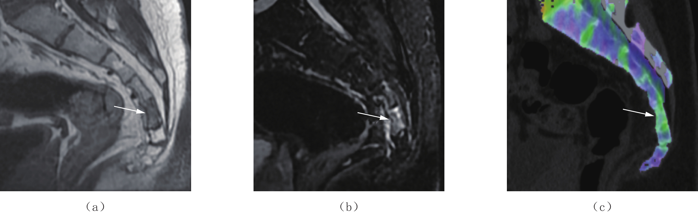
Purpose: The aim of this study was to evaluate the diagnostic performance of virtual non-calcium imaging using dual-energy computed tomography (CT) for acute sacrococcygeal injuries diagnosed by radiologists with varying levels of experience. Methods: A prospective study was employed and 29 patients presenting with acute coccygeal pain due to trauma between November 2021 and January 2024, involving a total of 174 vertebrae (145 sacral, 29 coccygeal), were enrolled. Both magnetic resonance imaging (MRI) and DECT data were acquired. Three radiologists with 1, 4, and 10 years of experience analyzed conventional CT and VNCa color-coded images, using MRI as the reference standard. Results: Following the application of VNCa imaging, the inter-rater agreement among the three radiologists significantly improved, with Kappa values increasing from 0.452, 0.615, and 0.735 to 0.775, 0.825, and 0.897, respectively. Diagnostic accuracy also notably increased to 92.5%, 94.3%, and 96.6% for the three radiologists, with no statistically significant differences observed among them. Quantitative analysis revealed an average VNCa CT value of (−84.2±20.3) HU for normal bone marrow and (−37.4±18.8) HU for bone marrow edema, showing a significant difference between the two. Using MRI as the reference standard, the area under the curve for distinguishing normal bone marrow from BME based on VNCa CT values was 0.962, with an optimal cutoff of −57.1 HU, yielding a sensitivity and specificity of 91.9% and 90.2%, respectively. Conclusion: DECT VNCa imaging significantly enhances the diagnostic efficacy of radiologists with varying levels of experience in acute coccygeal injury assessment, particularly benefiting less-experienced physicians. VNCa CT values contribute substantially to diagnostic accuracy in this context.
| [1] |
陈子锴, 袁荣霞, 方锐洁, 等. 郑氏提顶理筋手法结合润肠通便方对臀坐式骶尾椎骨折患者的临床疗效观察[J]. 中华中医药杂志, 2023, 38(10): 5087−5090.
CHEN Z K, YUAN R X, FANG R J, et al. Clinical efficacy observation of ZHENGs’ manipulation of lifting the top and regulating the tendons combined with Runchang Tongbian Recipe in patients with buttock sitting sacrococcygeal fracture[J]. China Journal of Traditional Chinese Medicine and Pharmacy, 2023, 38(10): 5087−5090. (in Chinese).
|
| [2] |
BARBER L A, KATSUURA Y, QURESHI S. Sacral fractures: A review[J]. HSS Journal, 2023, 19(2): 234-246. DOI: 10.1177/15563316221129607.
|
| [3] |
BURGHARDT A J, LINK T M, MAJUMDAR S. High-resolution computed tomography for clinical imaging of bone microarchitecture[J]. Clinical Orthopaedics and Related Research, 2011, 469(8): 2179−2193. DOI: 10.1007/s11999-010-1766-x.
|
| [4] |
BOOZ C, NÖSKE J, ALBRECHT M H, et al. Diagnostic accuracy of color-coded virtual noncalcium dual-energy CT for the assessment of bone marrow edema in sacral insufficiency fracture in comparison to MRI[J]. European Journal of Radiology, 2020, 129: 109046. DOI: 10.1016/j.ejrad.2020.109046.
|
| [5] |
HAN I H, CHIN D K, KUH S U, et al. Magnetic resonance imaging findings of subsequent fractures after vertebroplasty[J]. Neurosurgery, 2009, 64(4): 740-744. DOI: 10.1227/01.NEU.0000339120.41053.F1.
|
| [6] |
KAZAWA N. T2WI MRI and MRI-MDCT correlations of the osteoporotic vertebral compressive fractures[J]. European Journal of Radiology, 2012, 81(7): 1630−1636. DOI: 10.1016/j.ejrad.2011.04.052.
|
| [7] |
AKISATO K, NISHIHARA R, OKAZAKI H, et al. Dual-energy CT of material decomposition analysis for detection with bone marrow edema in patients with vertebral compression fractures[J]. Academic radiology, 2020, 27(2): 227−232. DOI: 10.1016/j.acra.2019.02.015.
|
| [8] |
PACHE G, KRAUSS B, STROHM P, et al. Dual-energy CT virtual noncalcium technique: Detecting posttraumatic bone marrow lesions—feasibility study[J]. Radiology, 2010, 256(2): 617-624. DOI: 10.1148/radiol.10091230.
|
| [9] |
GOSANGI B, MANDELL J C, WEAVER M J, et al. Bone marrow edema at dual-energy CT: A game changer in the emergency department[J]. Radiographics, 2020, 40(3): 859−874. DOI: 10.1148/rg.2020190173.
|
| [10] |
TIVNAN P, KALIAEV A, ANDERSON S W, et al. Utilization of a two-material decomposition from a single-source, dual-energy CT in acute traumatic vertebral fractures[J]. Frontiers in Radiology, 2023, 3: 1187449. DOI: 10.3389/fradi.2023.1187449.
|
| [11] |
LI Z, CHEN X, FANG H, et al. Diagnostic accuracy of dual-energy CT for bone marrow edema in patients with acute knee injury: A systematic review and meta-analysis[J]. Journal of Orthopaedic Surgery and Research, 2023, 18(1): 826. DOI: 10.1186/s13018-023-04151-3.
|
| [12] |
WALSTRA F E, HICKLE J, DUGGAN P, et al. Top-ten tips for dual-energy CT in MSK Radiology[J]. Seminars in Musculoskeletal Radiology, 2019, 23(4): 392−404. DOI: 10.1055/s-0039-1694756.
|
| [13] |
梁建超, 方义杰, 李文娟, 等. 双能量CT虚拟去骨图不同对比物质相对比值对膝关节创伤性骨髓水肿的诊断价值[J]. 中华放射学杂志, 2018, 52(1): 41−45. DOI: 10.3760/cma.j.issn.1005-1201.2018.01.009.
LIANG J C, FANG Y J, LI W J, et al. Diagnostic value of different related contrast material in dual-energy CT virtual noncalcium for detecting traumatic bone marrow edema in knee joint[J]. Chinese Journal of Radiology, 2018, 52(1): 41−45. DOI: 10.3760/cma.j.issn.1005-1201.2018.01.009. (in Chinese).
|
| [14] |
孔玲玲, 徐驰杰, 赵旻月, 等. 双能量CT虚拟单能量成像在鉴别脊柱急慢性压缩骨折中的应用价值[J]. CT理论与应用研究(中英文), 2021, 30(2): 209−216. DOI: 10.15953/j.1004-4140.2021.30.02.08.
KONG L L, XU C J, ZHAO M Y, et al. The application value of dual-energy CT virtual monoenergetic imaging in the differential diagnosis of acute and chronic spinal compression fractures[J]. CT Theory and Applications, 2021, 30(2): 209−216. DOI: 10.15953/j.1004-4140.2021.30.02.08. (in Chinese).
|
| [15] |
MENNEN A H M, BLOKLAND A S, MAAS M, et al. Imaging of pelvic ring fractures in older adults and its clinical implications-a systematic review[J]. Osteoporosis International, 2023, 34(9): 1549−1559. DOI: 10.1007/s00198-023-06812-9.
|
| [16] |
GRUNZ J P, SAILER L, LANG P, et al. Dual-energy CT in sacral fragility fractures: Defining a cut-off Hounsfield unit value for the presence of traumatic bone marrow edema in patients with osteoporosis[J]. BMC Musculoskeletal Disorders, 2022, 23(1): 724. DOI: 10.1186/s12891-022-05690-2.
|
| [17] |
PALM H G, LANG P, HACKENBROCH C, et al. Dual-energy CT as an innovative method for diagnosing fragility fractures of the pelvic ring: A retrospective comparison with MRI as the gold standard[J]. Archives of Orthopaedic and Trauma Surgery, 2020, 140(4): 473−480. DOI: 10.1007/s00402-019-03283-8.
|
| [18] |
中华医学会放射学分会, 中国医师协会放射医师分会, 安徽省影像临床医学研究中心. 能量CT临床应用中国专家共识[J]. 中华放射学杂志, 2022, 56(5): 476−487. DOI: 10.3760/cma.j.cn112149-20220118-00051.
Chinese Society of Radiology of Chinese Medical Association, Chinese Radiologist Association, Research Center of Clinical Medical Imaging of Anhui Province. China expert consensus on clinical application of multi-energy CT[J]. Chinese Journal of Radiology, 2022, 56(5): 476−487. DOI: 10.3760/cma.j.cn112149-20220118-00051. (in Chinese).
|
| [19] |
MAŁKIEWICZ A, DZIEDZIC M. Bone marrow reconversion: Imaging of physiological changes in bone marrow[J]. Polish Journal of Radiology, 2012, 77(4): 45−50. DOI: 10.12659/pjr.883628.
|
| [20] |
KELLOCK T T, NICOLAOU S, KIM S S Y, et al. Detection of bone marrow edema in nondisplaced hip fractures: Utility of a virtual noncalcium dual-energy CT application[J]. Radiology, 2017, 284(3): 922. DOI: 10.1148/radiol.2017161063.
|
| [21] |
SON W, PARK C, JEONG H S, et al. Bone marrow edema in non-traumatic hip: High accuracy of dual-energy CT with water-hydroxyapatite decomposition imaging[J]. European Radiology, 2020, 30(4): 2191−2198. DOI: 10.1007/s00330-019-06519-8.
|
| [22] |
CAVALLARO M, D'ANGELO T, ALBRECHT M H, et al. Comprehensive comparison of dual-energy computed tomography and magnetic resonance imaging for the assessment of bone marrow edema and fracture lines in acute vertebral fractures[J]. European Radiology, 2022, 32(1): 561−571. DOI: 10.1007/s00330-021-08081-8.
|
| [23] |
左天姿, 陈英敏, 贾秀川, 等. 双能CT虚拟去钙技术显示非创伤性股骨头坏死骨髓水肿的研究[J]. 实用放射学杂志, 2021, 37(4): 624−627, 632. DOI: 10.3969/j.issn.1002-1671.2021.04.027.
ZUO T Z, CHEN Y M, JIA X C, et al. Study on bone marrow edema of non-traumatic femoral head necrosis via dual-energy CT virtual noncalcium[J]. Journal of Practical Radiology, 2021, 37(4): 624−627, 632. DOI: 10.3969/j.issn.1002-1671.2021.04.027. (in Chinese).
|
| [24] |
JANG S W, CHUNG B M, KIM W T, et al. Nondisplaced fractures on hip CT: Added value of dual-energy CT virtual non-calcium imaging for detection of bone marrow edema using visual and quantitative analyses[J]. Acta Radiologica, 2019, 60(11): 1465−1473. DOI: 10.1177/0284185119831690.
|
| [25] |
LI M, QU Y, SONG B. Meta-analysis of dual-energy computed tomography virtual non-calcium imaging to detect bone marrow edema[J]. European Journal of Radiology, 2017, 95: 124−129. DOI: 10.1016/j.ejrad.2017.08.005.
|
| [1] | YAN Xin, ZHAO Jianhua. Research Progress of Pericoronary Adipose Tissue Radiomics Based on Coronary Computed Tomography Angiography[J]. CT Theory and Applications, 2024, 33(4): 531-538. DOI: 10.15953/j.ctta.2023.179 |
| [2] | HE Weihong, FANG Tingsong, FU Xi, LAO Meiling, XIAO Xiuyun. Risk Factors of Vulnerable Coronary Plaque Formation in Type 2 Diabetes[J]. CT Theory and Applications, 2023, 32(4): 523-529. DOI: 10.15953/j.ctta.2023.036 |
| [3] | ZHU Najun, FANG Xinxin, YIN Yijun, ZHOU Shuitian. Risk Factors of Plaque Progression in Patients with Angina Pectoris and Their Relationships with Coronary CT Angiography[J]. CT Theory and Applications, 2023, 32(2): 217-222. DOI: 10.15953/j.ctta.2022.219 |
| [4] | LIANG Yu, ZHANG Xiao-qin. Current State and Progress of Coronary Angiography in Multi-slice Spiral Computed Tomography Imaging Techniques[J]. CT Theory and Applications, 2016, 25(6): 725-735. DOI: 10.15953/j.1004-4140.2016.25.06.14 |
| [5] | HAN Hong-cheng. The Application of Multi Slice Spiral CT Angiography in Coronary Heart Disease[J]. CT Theory and Applications, 2015, 24(6): 843-848. DOI: 10.15953/j.1004-4140.2015.24.06.10 |
| [6] | YANG Chun-yu, SHEN Bi-xian, ZHAO Yue, HUANG Yin-ping, CHEN Sheng-ji, HUANG An-rong. Study on the Value of Dual Source CT Assessment of Correlation between Diabetes and Coronary Plaque[J]. CT Theory and Applications, 2014, 23(6): 913-921. |
| [7] | ZHAO Yue, SHEN Bi-xian, TAN Si-ping, YANG Chun-yu, CHEN Sheng-ji, HUANG An-rong. Study on the Value of Dual Source CT Assessment of Smoking and Coronary Plaque Correlation[J]. CT Theory and Applications, 2014, 23(4): 541-550. |
| [8] | TIAN Shu-ping, YANG Li, WANG Zhan-yu, WANG Shou-hai, WANG Zi-jun, ZHANG Yan-qun. The Application of “Magic Glass” Function in Coronary CTA[J]. CT Theory and Applications, 2012, 21(4): 721-726. |
| [9] | LIU Chao, ZHOU Xuan-ming, GONG Xiao-hong, CHEN Lun-gang. Clinical Significance of Evaluating Coronary Atherosclerotic Plaque with 64-Slice CT[J]. CT Theory and Applications, 2010, 19(2): 105-111. |
| [10] | XU Hui-min, HUO Jian-wei, LI Bao-ping, ZENG Qing-yu. Evaluation of Coronary Heart Disease using MDCT Coronary Angiography Combined with Ultrasound[J]. CT Theory and Applications, 2004, 13(4): 55-59. |
Supported by: Beijing Renhe Information Technology Co. Ltd 


 DownLoad: