Study on the Application of CTU and CTA Combined Imaging by Fractional Injection Combined of a Contrast Agent with Energy Imaging
-
摘要:
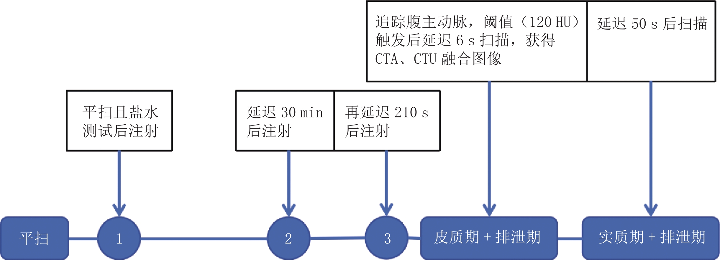
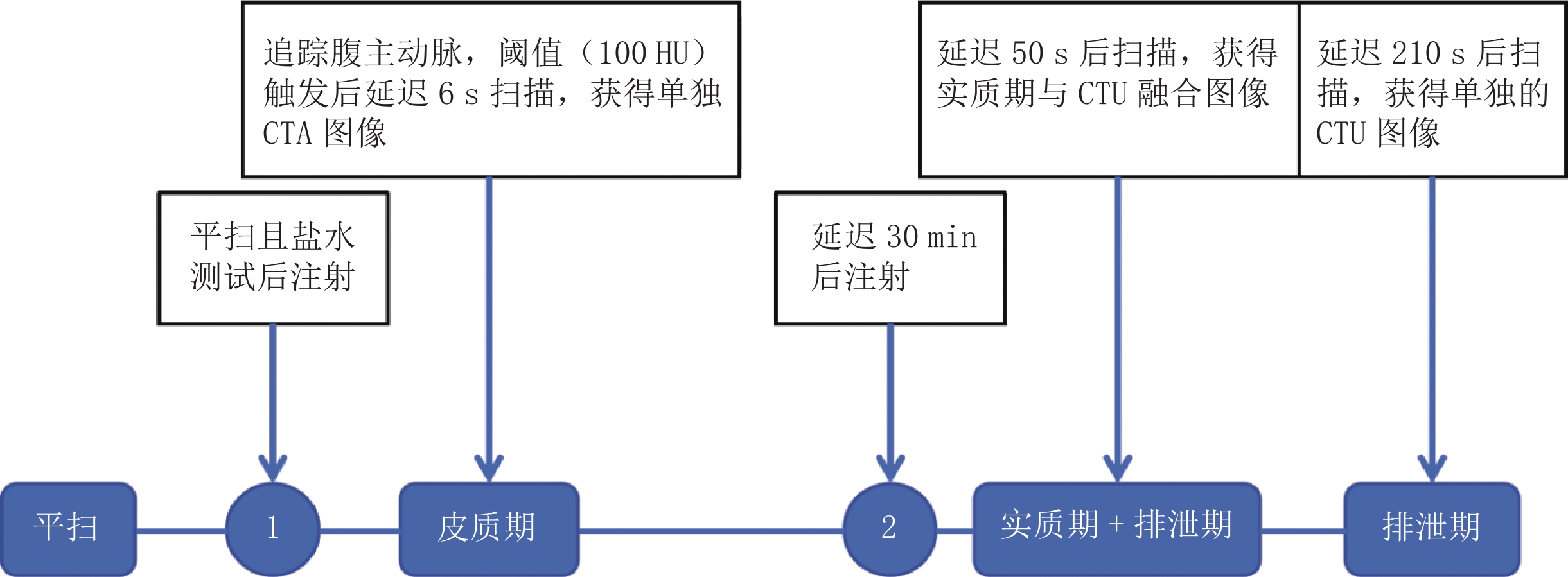
目的:探讨对比剂分次团注结合双层探测器光谱CT 50 keV虚拟单能量图像(VMI),在CT泌尿系成像(CTU)与主动脉CTA联合成像中的应用价值。方法:收集2024年3月至4月在首都医科大学附属北京同仁医院Philips IQon双层探测器光谱CT行CTU检查患者32例为试验组。使用对比剂分次团注及团注追踪技术;扫描后重建常规120 kVp混合能量图像为A1组;光谱重建获得50 keV VMI为A2组。收集2023年12月至2024年3月间在相同设备行CTU检查患者32例为对照组,使用对比剂分离团注技术及团注追踪技术;扫描后重建常规120 kVp混合能量图像为B组。客观评价包括:比较3组腹主动脉、双侧肾动脉的CT值、对比噪声比(CNR)、信噪比(SNR);比较输尿管起始部平均CT值和中下部平均CT值,并采用Kruskal-Wallis检验,采用Nemenyi检验进行两两比较。试验组与对照组的有效辐射剂量比较采用Wilcoxon检验。主观评价由两名高年资影像医师分别对3组CTA及CTU图像进行4分法和3分法评价,两名医师主观评分的一致性采用Kappa检验。结果:客观评价指标中,A2组分别与A1组和B组比较,均有统计学差异;A1组与B组仅双侧肾动脉SNR有统计学差异。两组有效辐射剂量有统计学差异,试验组较对照组降低15.1%。3组CTA、CTU图像主观评价无统计学差异;两名医师对CTA图像评分结果一致性非常好,对CTU图像评分结果一致性好。结论:利用对比剂分次团注行CTU检查时,获得泌尿系及主动脉同时显影的融合图像,不仅具有明确的解剖关系及相同的图像质量,而且降低有效辐射剂量;光谱重建获得的50 keV虚拟单能量图像,优化CTU及CTA图像质量,具有临床价值。
Abstract:Objective: This study aimed to investigate the application value of fractional bolus injection of a contrast agent combined with double-layer detector spectral CT 50 keV virtual monoenergetic imaging (VMI) in combined imaging of computed tomography urography (CTU) and aortic CT angiography (CTA). Methods: The experimental group included 32 patients who underwent spectral CTU with a Philips IQon dual-layer detector at Beijing Tongren Hospital, Capital Medical University, between March and April 2024. Using fractional bolus injection of a contrast agent and bolus tracking technology, the conventional 120 kVp mixed energy image was reconstructed after scanning for group A1; the 50 keV VMI obtained by spectral reconstruction was group A2. The control group included 32 patients who underwent CTU examination between December 2023 and March 2024 using the same equipment, and the contrast agent split bolus and group injection tracking techniques were used. After scanning, the conventional 120 kVp mixed energy image was reconstructed for group B. The objective evaluation included the CT value, contrast-to-noise ratio (CNR), and signal-to-noise ratio (SNR) of the abdominal aorta and bilateral renal arteries, which were compared among the three groups. The Kruskal–Wallis and Nemenyi tests were used to compare the average CT values of the initial, middle, and lower parts of the ureter. Wilcoxon test was used to compare the effective radiation dose between the experimental and control groups. Subjective evaluation was performed by two senior radiologists on the three groups of CTU and CTA images using the 4- and 3-point methods, respectively. The consistency of the subjective scores of the two radiologists was analyzed using the Kappa test. Results: Among the objective evaluation indices, there were statistically significant differences among groups A2, A1, and B. There was a significant difference in the SNR of the bilateral renal arteries between groups A1 and B. The effective radiation dose of the experimental group was 15.1% lower than that of the control group, which was significantly different. There was no significant difference in the subjective evaluation of the CTA and CTU images among the three groups. The consistency of the CTA and CTU image scoring results between the two physicians was excellent and good, respectively. Conclusion: If CTU examination was performed by fractional injection of a contrast agent, fusion images of the urinary system and aorta were obtained, which not only had a clear anatomical relationship and the same image quality, but also reduced the effective radiation dose. The 50keV virtual monoenergetic image obtained by spectral reconstruction optimizes the image quality of CTU and CTA and has clinical value.
-
COVID-19是一种具有高度传染性的疾病,对全球人民的生命和健康造成了很大威胁。2020年1月30日世界卫生组织(WHO)宣布本次疫情为全球突发公共卫生事件,并提高了风险等级。截至2023年02月03日,全球已报道超过7.54亿病例感染、超过681万人死亡[1]。胸部CT的一些影像学特征对COVID-19的诊断有意义[2]。
COVID-19的起病、发展、转归时期的临床指标及胸部CT的影像学特征是一个动态演变的过程,本文通过比较分析处于不同发病时间的COVID-19患者的临床指标与多种胸部CT影像特征,总结处于不同病程的COVID-19患者临床指标与多种胸部CT影像特征的发展规律,对临床医生的诊断与治疗提供帮助。
1. 材料与方法
1.1 研究人群
收集2022年11月至2023年1月期间首都医科大学附属北京世纪坛医院发热门诊收治的161例新型冠状病毒抗原阳性同时胸部CT显示肺部感染阳性的病例,其中男91例(56.5%),女70例(43.5%),年龄26~98岁,平均年龄(68.5±15.8)岁;病程1~35 d,平均病程(9.1±5.6) d;按首次发热时间与CT检查时间间隔不同分为两组(<10 d组,≥10 d组)。
本研究纳入的临床指标包括临床症状(发热、憋气、咳嗽、咳痰、咽痛、肌痛)、实验室指标(C反应蛋白、白细胞、淋巴细胞、单核细胞、中性粒细胞计数)。
1.2 CT扫描方法
采用32排北京SINOVISION CT Insitum-CT 338机型。扫描参数:探测器宽度16 cm,螺距1.0,管电压120 kV,管电流150 mAS,重建层厚肺窗1.5 cm,层间距1.5 cm,纵隔窗5 mm,层间距5 mm,重建矩阵512×512,FOV 380~450。扫描范围肺尖至肺底。
1.3 影像分析
由两名有经验的放射科医生分别阅片,对161例病例的胸部CT影像特征进行评价,如意见不一致,由高级别医生决定。评价的CT影像特征包括:周围(胸膜下、胸膜内)、中央(血管周、血管外)、混合分布、上肺为主,下肺为主,斑片、大片、束带、铺路石征、空气支气管征、空泡征、蜂窝、边界不规则、病灶内条索、纤维索条、牵拉支扩、反晕征、煎蛋征、胸膜尾征、胸膜下线、胸膜下栅栏、胸膜增厚、叶间裂增厚、胸腔积液。
其中周围分布中的胸膜下分布指病变与胸膜相连;胸膜内分布指病变与胸膜间存在胸膜下黑线或胸膜下黑带即与胸膜间存在未受累区;中央分布中的血管周指病变分布在血管周围、紧邻血管;血管外指病变与血管间存在未受累区。
1.4 统计学方法
采用SPSS 26.0软件,根据发病与CT检查时间间隔将患者分为两组:<10 d组和≥10 d组,比较两组患者相关的临床指标与影像学征象。选择10 d作为分组的依据:有研究发现在轻度和中度COVID-19患者中,传染性病毒可以在症状发作后持续一周或更长时间,并随着时间的推移而下降,在症状出现后10 d,病毒培养的可能性降至6%[3]。
本研究数据均为非正态资料,以中位数及四分位数表示,组间比较采用曼-惠特尼检验、分类变量使用卡方检验或费舍尔精确检验。P<0.05表示差异有统计学意义。
2. 结果
两组COVID-19患者中,临床症状、实验室化验指标、CT影像学征象存在统计学差异。临床症状中咽痛和肌痛在两组间的差异有统计学意义,咽痛在<10 d组发生几率更大,肌痛在≥10 d组发生几率更大;而发热、憋气、咳嗽、咳痰两组间未见明确统计学意义(表1)。
表 1 不同病程患者组临床症状占比情况Table 1. Proportion of clinical symptoms in different disease course groups临床指标 组别 统计检验 <10 d ≥10 d $Z/\chi^{2}$ P 年龄($M(Q_1,Q_3)$) 69(59,82) 70(59,79) -0.328 0.743 性别(男,例(%)) 52(56.5) 39(56.5) 0.000 1.000 发热/(例(%)) 92(100.0) 69(100.0) — — 憋气/(例(%)) 13(14.1) 14(20.3) 1.072 0.301 咳嗽/(例(%)) 45(48.9) 37(53.6) 0.112 0.738 咳痰/(例(%)) 84(91.3) 64(92.8) 0.350 0.554 咽痛/(例(%)) 43(46.7) 19(27.5) 6.140 0.013 肌痛/(例(%)) 3(3.3) 9(13.0) 5.470 0.019 在实验室指标中,C反应蛋白的差异存在统计学意义,C反应蛋白在<10 d组数值更高淋巴细胞计数更低;而白细胞、中性粒细胞和单核细胞计数在两组间未见明确统计学差异(表2)。
表 2 不同病程患者组实验室指标对比情况Table 2. Comparison of laboratory indicators in different disease course groups实验室指标($M(Q_1,Q_3)$) 组别 统计检验 <10 d ≥10 d Z P C反应蛋白/(mg/L) 32.71(11.52,67.57) 15.70(2.88,50.93) -2.761 0.006 白细胞/(×109/L) 6.23(4.82,7.74) 6.99(5.10,8.29) -1.570 0.116 淋巴细胞/(×109/L) 1.31(0.99,1.69) 1.78(1.03,2.41) -2.979 0.003 单核细胞/(×109/L) 0.35(0.44,0.61) 0.47(0.37,0.59) -0.297 -0.766 中性粒细胞/(×109/L) 4.09(3.01,5.89) 4.22(3.19,5.90) -0.478 0.632 在CT影像特征方面,<10 d组患者存在血管周、混合分布、大片(图1(a))、空气支气管征(图1(a))的比例更高,≥10 d组患者存在边界不规则、病灶内索条(图1(d)和(e))、反晕征(图1(b)和(f))、胸膜尾征(图1(d))、胸膜下线(图1(c))、胸膜下栅栏(图1(c))的比例更高,差异有统计学意义。而胸膜下、胸膜内、血管外、上肺为主、斑片、束带状、实变相关征象、铺路石征、牵拉支扩、空泡征、煎蛋征、蜂窝、胸膜增厚、叶间裂增厚、胸腔积液出现占比在两组间差异没有统计学意义(表3)。
表 3 不同病程患者组病灶各类影像征象占比情况Table 3. Proportion of various imaging signs in the lesions of patients with different disease course影像指标 组别 统计检验 <10 d(92例) ≥10 d(69例) $\chi^{2} $ P 分布特征/(例(%)) 周围 88(95.7) 68(98.6) 1.101 0.294 胸膜下 67(72.8) 49(71.0) 0.064 0.800 胸膜内 85(92.4) 66(95.7) 0.720 0.396 中央 74(80.4) 45(65.2) 4.735 0.030 血管周 74(80.4) 43(62.3) 6.515 0.011 血管外 10(10.9) 8(11.6) 0.021 0.885 混合 71(77.2) 42(60.9) 5.009 0.025 分布优势/(例(%)) 上肺为主 10(10.9) 6(8.7) 0.208 0.648 下肺为主 41(44.6) 43(62.3) 3.846 0.050 病变形态/(例(%)) 斑片状 81(88.0) 56(81.2) 1.473 0.225 大片状 56(60.9) 25(36.2) 9.574 0.002 束带状 39(42.4) 34(49.3) 0.754 0.385 实变相关征象/(例(%)) 42(45.7) 29(42.0) 0.210 0.647 铺路石征 56(60.9) 35(50.7) 1.651 1.199 空气支气管征 69(75.0) 32(46.4) 13.817 0.000 空泡征 52(56.5) 38(55.9) 0.006 0.936 机化纤维化征象/(例(%)) 蜂窝 9(9.8) 9(13.0) 9.414 0.516 边界不规则 40(43.5) 44(63.8) 6.505 0.011 病灶内索条 22(23.9) 37(53.6) 14.991 0.000 纤维索条 61(66.3) 51(73.9) 1.078 0.299 牵拉支扩 50(54.3) 45(65.2) 1.926 0.165 反晕征 30(32.6) 37(53.6) 7.166 0.007 煎蛋征 46(50.0) 42(60.9) 1.880 0.170 胸膜尾征 38(41.3) 44(63.8) 7.961 0.005 胸膜下线 21(22.8) 34(49.3) 12.264 0.000 胸膜下栅栏 7(7.6) 26(37.7) 21.881 0.000 胸膜病变/(例(%)) 胸膜增厚 68(73.9) 56(81.2) 1.170 0.279 叶间裂增厚 14(15.2) 17(24.6) 2.251 0.134 胸腔积液 3(3.3) 3(4.3) 0.130 0.719 3. 讨论
3.1 不同病程COVID-19患者的临床表现和实验室指标分析
研究显示,COVID-19常见的临床症状主要为发热、咳嗽,咳痰、畏寒、头痛、咽痛、胸闷气促、流涕、肌痛、乏力、腹痛等,有研究认为咽痛更多见于轻症,而肌痛在重症患者中更多见[4];在本研究中,咽痛在<10 d组更常见,而肌痛在≥10 d组更常见,结果支持了既往研究。
C反应蛋白(CRP)已被公认为全身炎症和严重感染的标志物[5]。作为一种急性期反应物,CRP与宿主细胞病原体和膜中的磷酸胆碱结合,增强吞噬作用、促进清除,此外,CRP与配体结合后还可以效地激活补体系统[6]。C反应蛋白水平的升高与全身炎症反应和COVID-19患者的严重程度紧密相关[7-8]。本研究重症患者较少,主要为轻型和中型患者,C反应蛋白在病毒感染的急性期升高,而随着病程时间延长,炎症逐渐吸收,C反应蛋白呈下降趋势,符合炎症发展的规律。
有文献报道,淋巴细胞在COVID-19急性期表现为数量减少,在恢复期其数量逐渐升高至正常水平[9-10]。在本研究中,相对于≥10 d组而言,<10 d组淋巴细胞计数下降更明显,提示随病程的延长,肺部感染逐渐吸收好转,本研究与文献一致。
3.2 不同病程的COVID-19患者CT征象分析
研究显示,新型冠状病毒利用血管紧张素转化酶-2(ACE2)作为细胞受体后,首先引起肺间质损伤、随后伴有实质的变化[2,11-12]。有文献基于尸检结果发现,新型冠状病毒相关的肺部病理改变以3个阶段的弥漫性肺泡损伤(DAD)为特征,即渗出、增生和纤维化[13]。大片影(>3 cm)是肺部炎症渗出、实变的表现,在<10 d组占比更高,而随着病程延长,炎症渗出病变逐渐吸收,大片影随之减少。血管周和混合分布说明炎症累及范围较广,在<10 d组占比更高,说明随着病程延长,炎症范围减小。
在影像学征象方面,本研究显示不同病程的COVID-19多个征象存在明显的统计学差异。支气管充气征(air bronchogram)是指在不透明背景下可以看到充气支气管影,据报道是COVID-19的CT表现[2,10]。支气管充气征通常在大片影背景里存在,这就可以解释支气管充气征发生规律和大片影相同。反晕征(reversed halo sign),又称环礁征(atoll sign),表现为中心是密度较低的磨玻璃密度影,周围是新月形或环形的高密度实变影[2,14]。本次研究中,≥10 d组反晕征发生机率更多,可能是由于随着病程延长、疾病的发展,围绕GGO发生实变、增殖、机化,或是病变吸收而在中心留下相对低密度[15-16]。
在COVID-19中观察到纤维化或纤维索条的CT表现。有研究显示[17],17% 的COVID-19患者存在纤维条索。纤维化可能是由于肺慢性炎症或增殖性疾病愈合期间细胞成分逐渐被瘢痕组织取代而形成的。本研究中,纤维索条在两组间的差异未见明确统计学差异,原因可能如下:
①本研究中纳入的患者多为老年 COVID-19患者,其本身存在基础性纤维索条改变;②纤维索条在肺部CT中是较为常见的征象,炎症、结核等很多原因都可以导致,同时,纤维索条在10 d后出现更可能是疾病转归的一种表现,而一次检查无法评价索条是否为陈旧性病变。
基于以上的原因,本研究引入病灶内索条的概念,在≥10 d组病灶内索条占比更多,更能反映病变内部出现了纤维化的表现。
胸膜下线、胸膜下栅栏、胸膜尾征的出现提示出现肺部纤维化改变。对于以上几个COVID-19患者的CT相关影像学征象,目前研究较少。本研究中≥10 d组出现胸膜下线、胸膜下栅栏、胸膜尾征的机率更大,提示随着病程延长、疾病的发展,肺部渗出、实变病灶逐渐吸收,出现了肺部纤维化改变。
目前,纤维化与患者预后之间的关系值得商榷。部分研究认为纤维化的存在表明疾病状态稳定的COVID-19患者预后良好[15]。然而,也有研究显示纤维化可能表明COVID-19预后不良,认为纤维化征象提示其随后可能进展到高峰期或导致肺间质纤维化疾病[16-18]。
本研究存在部分局限性:①本研究纳入患者多为轻中症患者,下一步可增加重症患者的比例进行研究;②本研究未纳入肺功能成像指标;③本研究仅分析早期纤维化相关征象的差异,可进一步纳入长期的COVID-19患者进行研究。
综上所述,不同病程的COVID-19患者不仅在临床症状、实验室指标方面存在明显差异,在CT图像上病变的分布、形态、特殊征象方面均存在显著差异,认识这些差异存在的规律,可以帮助临床医生更好地诊断和治疗COVID-19。需要特别指出的是,本研究发现在发病10 d后,肺部机化和纤维化的影像学征象占比更高、对比<10 d组有显著差异,提示临床医生在发病10 d或更早时间使用抗纤维化药物是有一定必要性的。
-
图 3 不同扫描方法对结石显示的MIP图像及A2组、A1组VR图像
注:(a)为B组CTU单独显影的MIP像(男,48岁,BMI为22.68),显示左输尿管结石(红圈处),(b)为A2组CTA、CTU同时显影的MIP图像(男,43岁,BMI为25.26),显示右侧输尿管与髂动脉交界处结石(红圈处);(c)、(d)分别为A2组、A1组同一患者VR图像(男,25岁,BMI为23.18);试验组采用对比剂分次团注方式,达到CTA、CTU同时显影的效果,显示由于髂动脉压迫导致输尿管狭窄(红圈处),A2组CT值高,显示细节丰富。
Figure 3. MIP images of different scanning methods for the display of stones and VR images of group A2 and group A1
表 1 不同体重使用对比剂总量、每段对比剂用量及注射流率
Table 1 The total amount of contrast agent, amount of contrast agent per segment, and injection flow rate used according to body weights
体重/kg 总对比剂量/mL 每段对比剂用量/mL 注射流率/(mL/s) 60 72 24 2.4 70 84 28 2.8 80 96 32 3.2 90 108 36 3.6 表 2 两组一般情况比较(
$ \bar{x}\pm s$ )Table 2 Comparison of two general situation groups (
$\bar{x}\pm s $ )项目 组别 统计检验 对照组 试验组 统计值 P 男/女 23/9 23/9 0.000 1.000 年龄/岁 53±17 55±16 0.021 0.885 BMI/(kg/m2) 24.76±3.04 24.46±2.89 0.112 0.739 表 3 3组图像质量客观评价(M(Q1,Q3))
Table 3 Objective evaluation of three image quality groups (M(Q1, Q3))
项目 组别 统计检验 A1 A2 B 统计值 P 例数 32 32 32 - - 腹主动脉 CT值/HU 257.80(221.30,294.00) 518.95(432.40,598.95) 295.60(260.55,310.40) 65.937 <0.001 CNR 11.96(10.28,15.36) 31.31(28.04,40.22) 10.76(9.69,12.46) 64.201 <0.001 SNR 11.73(10.24,16.05) 27.50(22.52,35.90) 9.90(8.38,11.08) 66.155 <0.001 左肾动脉 CT值/HU 233.20(199.45,266.75) 458.90(383.40,531.35) 269.85(239.05,291.20) 65.815 <0.001 CNR 11.46(8.92,13.24) 30.50(25.05,37.48) 8.95(7.51,10.74) 66.008 <0.001 SNR 12.25(9.59,16.85) 20.76(15.95,28.04) 8.13(6.74,10.77) 46.748 <0.001 右肾动脉 CT值/HU 238.50(193.30,256.15) 449.55(374.80,544.55) 261.50(239.05,284.80) 59.555 <0.001 CNR 11.36(8.633,14.23) 33.33(24.98,35.14) 9.15(7.30,10.05) 67.002 <0.001 SNR 12.04(9.73,17.55) 21.51(13.36,27.54) 7.52(6.45,9.16) 49.132 <0.001 平均CT值/HU 输尿管起始部 428.42(296.30,610.72) 778.70(567.45, 1112.50 )427.83(334.68,668.68) 24.193 <0.001 输尿管中下部 417.50(277.77,666.50) 784.58(589.95, 1020.45 )470.88(334.28,564.17) 25.260 <0.001 同层面膀胱内近腹部CT值与中部CT值比 0.75 0.66 0.54 - - 左右髂动脉平均CT值/HU 258.58(216.30,296.45) 500.55(409.08,564.60) - 70.196 <0.001 腹主动脉CT值/HU 257.80(221.30,294.00) - 295.60(260.55,310.40) 表 4 两名医师对3组CTA、CTU图像主观评分
Table 4 The subjective scores of CTA and CTU images of the three groups scored by two physicians
项目 组别 医师1/例 医师2/例 4分 3分 2分 1分 4分 3分 2分 1分 CTA图像 A1 - 32 0 0 - 32 0 0 A2 - 32 0 0 - 32 0 0 B - 31 1 0 - 32 0 0 CTU图像 A1 19 8 5 0 12 14 6 0 A2 19 8 5 0 13 13 6 0 B 18 11 3 0 13 14 5 0 表 5 两组有效辐射剂量比较
Table 5 Comparison of effective radiation doses between the two groups
组别 对照组 试验组 统计值 P ED/mSv 33.59(27.44,45.27) 28.81(24.96,33.37) -2.041 0.041 -
[1] 中华医学会影像技术分会, 中华医学会放射学分会. CT检查技术专家共识[J].中华放射学杂志, 2016, 50(12): 916-928. DOI: 10.3760/cma.j.issn.1005-1201.2016.12.004. [2] 丁宁, 王彬, 许开元, 等. 多排螺旋CT同步肾动脉和肾盂输尿管显影在腔镜手术中的临床价值[J]. 中华腔镜泌尿外科杂志(电子版), 2011, 5(3): 213−217. DOI: 10.3877/cma.j.issn.1674-3253.2011.03.011. DING N, WANG B, XU K Y, et al. Clinical value of multiple-slice spiral CT synchronous imaging of renal arteries, renal pelves and ureters in endoscopic surgeries[J]. Chinese Journal of Endourology (Electronic Edition), 2011, 5(3): 213−217. DOI: 10.3877/cma.j.issn.1674-3253.2011.03.011. (in Chinese).
[3] 张小平, 李兴国, 刘承杏, 等. 肾盂输尿管连接部与肾血管关系及其临床意义[J]. 中国临床解剖学杂志, 2006, 24(2): 153−156. DOI: 10.3969/j.issn.1001-165X.2006.02.012. ZHANG X P, LI X G, LIU C X, et al. The relationship between ureteropelvic junction and the vessels of kidney and its clinical significance[J]. Chinese Journal of Clinical Anatomy, 2006, 24(2): 153−156. DOI: 10.3969/j.issn.1001-165X.2006.02.012. (in Chinese).
[4] 中华放射学杂志双层探测器光谱CT临床应用协作组. 双层探测器光谱CT临床应用中国专家共识(第一版)[J]. 中华放射学杂志, 2020, 54(7): 635−643. DOI: 10.3760/cma.j.cn112149-20200513-00679. Chinese Journal of Radiology Cooperative Group of Clinical Application of Dual-layer Spectral Detector CT. China expert consensus on clinical application of dual-layer spectral detector CT[J]. Chinese Journal of Radiology, 2020, 54(7): 635−643. DOI: 10.3760/cma.j.cn112149-20200513-00679. (in Chinese).
[5] 范瑜, 殷焱, 王文晶, 等. 分离团注技术在CTU检查中的优势[J]. 医学影像学杂志, 2012, 22(9): 1547−1549. DOI: 10.3969/j.issn.1006-9011.2012.09.048. FAN Y, YIN Y, WANG W J, et al. Advantage of separate bolus injection technique in CT urograph[J]. Journal of Medical Imaging, 2012, 22(9): 1547−1549. DOI: 10.3969/j.issn.1006-9011.2012.09.048. (in Chinese).
[6] 贾紫婷. 多模态医学图像融合的关键技术研究[J]. 计算机时代, 2020, (7): 4−6, 11. DOI: 10.16644/j.cnki.cn33-1094/tp.2020.07.002. JIA Z T. Overview of key technologies of multimodal medical image fusion[J]. Computer Era, 2020(7): 4−6, 11. DOI: 10.16644/j.cnki.cn33-1094/tp.2020.07.002. (in Chinese).
[7] 顾恒乐, 聂生东. 多模医学图像配准和融合方法及其临床应用进展[J]. 中华放射肿瘤学杂志, 2016, 25(8): 902−906. DOI: 10.3760/cma.j.issn.1004-4221.2016.08.024. GU H L, NIE S D. Research advance in multi-modality medical image registration and fusion methods and their clinical application[J]. Chinese Journal of Radiation Oncology, 2016, 25(8): 902−906. DOI: 10.3760/cma.j.issn.1004-4221.2016.08.024. (in Chinese).
[8] 刘云福, 康天良, 张永县, 等. 两段式对比剂注射结合团注跟踪技术在肺动脉CTA检查中的应用研究[J]. CT理论与应用研究, 2023, 32(4): 531−538. DOI: 10.15953/j.ctta.2023.075. LIU Y F, KANG T L, ZHANG Y X, et al. Study of two-stage injection of contrast agent in combination with bolus tracking technique in computed tomography pulmonary angiography[J]. CT Theory and Applications, 2023, 32(4): 531−538. DOI: 10.15953/j.ctta.2023.075. (in Chinese).
[9] KEKELIDZE M, DWARKASING R S, DIJKSHOORN M L, et al. Kidney and urinary tract imaging: Triple-bolusmultidetector CT urography as a one-stop shop--protocol design, opacification, and image quality analysis[J]. Radiology, 2010, 255(2): 508-516. DOI: 10.1148/radiol.09082074.
[10] 卢向彬, 周鑫, 史浩. CTA、CTU联合应用在泌尿系统疾病中的应用价值研究[J]. 医学影像学杂志, 2013, 23(7): 1089−1091, 1110. DOI: 10.3969/j.issn.1006-9011.2013.07.033. LU X B, ZHOU X, SHI H. Value of CTA combined with CTU in the urogenital system diseases[J]. Journal of Medical Imaging, 2013, 23(7): 1089−1091, 1110. DOI: 10.3969/j.issn.1006-9011.2013.07.033. (in Chinese).
[11] 姜文龙, 杨艺, 吴志斌, 等. 结合能谱纯化与ADMIRE技术的一站式泌尿系CTA和CTU成像1例[DB/OL]. 中国临床案例成果数据库, 2023, 5(1): E02343-E02343. DOI: 10.3760/cma.j.cmcr.2023.e02343. JIANG W L, YANG Y, WU Z B, et al. A case of one-stop urological CTA and CTU imaging in combination with energy spectrum purification and ADMIRE technology was studied[DB/OL]. Chinese Medical Case Repository, 2023, 5(1): E02343-E02343. DOI:10.3760/cma.j.cmcr.2023.e02343. (in Chinese).
[12] 王鹤, 孙晓伟, 王继琛, 等. 正常集合系统分次团注双期与传统单次团注多期CT泌尿系造影的比较[J]. 中国医学影像技术, 2009, 25(11): 2076−2080. DOI: 10.3321/j.issn:1003-3289.2009.11.039. WANG H, SUN X W, WANG J C, et al. Comparasion of split-bolus2-phase CT urography and classical single bolus CT urography in normal collecting system[J]. Chinese Journal of Medical Imaging Technology, 2009, 25(11): 2076−2080. DOI: 10.3321/j.issn:1003-3289.2009.11.039. (in Chinese).
[13] KAREN C, FIONA C, LEJLA A, et al. CT urography: How to optimize the technique[J]. Abdominal Radiology, 2019, 44(12): 3786−3799. DOI: 10.1007/s00261-019-02111-2.
[14] 中华医学会放射学分会对比剂安全使用工作组. 碘对比剂使用指南(第2版)[J]. 中华医学杂志, 2014, 94(43): 3363−3369. DOI: 10.3760/cma.j.issn.0376-2491.2014.43.003. [15] WANG Z Q, SONG Y P, A G, et al. Role of hydration in contrast-induced nephropathy in patients who underwent primary percutaneous coronary intervention[J]. International Heart Journal, 2019, 60(5): 1077-1082. DOI: 10.1536/ihj.18-725.
[16] 中华医学会放射学分会心胸学组. 主动脉夹层CT血管成像标注专家共识[J]. 中华放射学杂志, 2024, 58(3): 250−257. DOI: 10.3760/cma.j.cn112149-20230719-00010. Cardiothoracic Group of Chinese Society of Radiology of Chinese Medical Association. Expert consensus on CT angiography annotation of aortic dissection[J]. Chinese Journal of Radiology, 2024, 58(3): 250−257. DOI: 10.3760/cma.j.cn112149-20230719-00010. (in Chinese).
[17] RAJIAH P, ABBARA S, HALLIBURTON S S. Spectral detector CT for cardiovascular applications[J]. Diagnostic and Interventional Radiology, 2017, 23(3): 187−193. DOI: 10.5152/dir.2016.16255.
[18] 中华医学会放射学分会, 中国医师协会放射医师分会, 安徽省影像临床医学研究中心. 能量CT临床应用中国专家共识[J]. 中华放射学杂志, 2022, 56(5): 476−487. DOI: 10.3760/cma.j.cn112149-20220118-00051. Chinese Society of Radiology of Chinese Medical Association, Chinese Radiologist Association, Research Center of Clinical Medical Imaging of Anhui Province. China expert consensus on clinical application of multi-energy CT[J]. Chinese Journal of Radiology, 2022, 56(5): 476−487. DOI: 10.3760/cma.j.cn112149-20220118-00051. (in Chinese).
[19] MICHAELA C, MAURIZIO C, NICOLO' R, et al. Computed tomography urography: State of the art and beyond[J]. Tomography, 2023, 9(3): 909−930. DOI: 10.3390/tomography9030075.
[20] 谢馥淳, 侯阳. 双层探测器光谱CT在外周血管成像中的应用进展[J]. 中华放射学杂志, 2021, 55(12): 1341−1344. DOI: 10.3760/cma.j.cn112149-20210816-00760. XIE F C, HOU Y. Application advantages of dual-layer spectral detector CT in peripheral vessels[J]. Chinese Journal of Radiology, 2021, 55(12): 1341−1344. DOI: 10.3760/cma.j.cn112149-20210816-00760. (in Chinese).


 下载:
下载:


