The Value of Subtraction Computed Tomography in Evaluating the Enhancement Degree of Thoracic Lesions
-
摘要:
目的:探讨CT肺减影成像在评价胸部占位性病变强化程度中的应用价值。方法:回顾性收集因胸部占位行胸部增强CT检查的患者,所有患者均采用CT肺减影成像序列扫描。经减影后处理得到动、静脉期的病灶碘分布彩色融合图。测量并计算动、静脉期无减影CT的强化净增值(∆Hu,∆Hu=强化后CT值-平扫CT值);病灶强化程度分为轻度强化(∆Hu<20 HU)、中度强化(∆Hu为20~40 HU)和显著强化(∆Hu>40 HU)。对CT肺减影伪彩图进行视觉评估,依据彩图的色阶将病灶碘分布程度分为轻度、中度、显著强化。随后测量病灶动脉期、静脉期CT肺减影图像的灌注值(∆Husub)。采用McNemar检验和kappa检验分别评估动、静脉期∆Hu等级和减影彩图视觉评估碘分布等级的差异和一致性。采用配对t检验对比动、静脉期普通CT的∆Hu和减影CT的∆Husub之间的差异。结果:共纳入34位患者(37个病灶)。3位观察者所测量数据的一致性良好。动、静脉期∆Hu分级和CT减影彩图视觉评估病灶碘分布等级之间无显著差异,且两者一致性好。动、静脉期无减影CT的∆Hu和有减影CT的∆Husub之间的差异均无统计学意义。结论:CT肺减影成像在不延长扫描时长的前提下,简化阅片时对病灶进行测量和计算的步骤,有助于提高诊断效率。
Abstract:Objective: This study aimed to explore the clinical use of the chest computed tomography (CT) subtraction technique for evaluating the degree of chest mass enhancement. Methods: Patients with suspected chest masses who underwent contrast-enhanced chest CT were retrospectively enrolled. Contrast-enhanced CT used was performed using lung subtraction. Subtraction images were reconstructed, and color fusion images of the iodine distribution in the chest masses in the arterial and venous phases were obtained after subtraction. The net enhancement value (∆Hu) was measured and calculated. The enhancement degree were divided into mild (∆Hu < 20 HU), moderate (∆Hu 20~40 HU), and significant (∆Hu > 40 HU). The CT subtraction iodine maps were visually evaluated. The net perfusion value (∆Husub) of CT lung subtraction iodine maps in arterial phase and venous phase was measured. McNemar and kappa tests were used to evaluate the difference and consistency of ∆Hu and ∆Husub degrees, respectively. The paired t test was used to compare the differences between ∆Hu and ∆Husub. Results: This study included 34 patients (37 lesions). Data measured by the three observers were in good agreement. The ∆Hu and ∆Husub degrees had no significant difference, but had good consistency. No significant differences were observed between ∆Hu and ∆Husub in the arterial and venous phases. Conclusion: Lung subtraction CT imaging can simplify the steps of image reading and report writing by reducing the measurement and calculation times for lung lesions without increasing scanning time, which contributes to improving the efficiency of diagnosis.
-
Keywords:
- computed tomography scanner /
- subtraction technique /
- chest mass /
- contrast media
-
胸部占位性病变在临床中十分常见,对病变的早期诊断、性质评估、术前分期是临床诊疗管理的关键[1]。胸部增强计算机断层扫描(computer tomography,CT)是诊断胸部占位性病变的常规检查手段[2−3],不仅可以提供病灶的大小、形态、密度、毗邻关系等影像特征信息,还可以通过病变的血供特征,为良、恶性胸部占位性病变提供鉴别依据[4−5]。因此胸部医学影像检查已成为目前医学影像科工作量最大的组别之一,面对激增的胸部增强CT诊断工作量,如何在保证诊断准确率和诊断报告书写质量的同时,简化阅片步骤,提升工作效率,是胸部影像诊断医师十分关心的问题。
CT肺减影技术可在扫描机自带软件上对图像进行处理,简化了流程,提高了工作效率[6]。然而,CT肺减影图测得的病灶强化值能否替代普通增强CT图计算的强化净增值尚不明确,且CT肺减影技术在胸部占位性病变中的研究较少。因此,本研究旨在对比肺减影CT和普通增强CT在评估胸部占位性病变强化程度中的差异,以探讨CT肺减影技术是否可以在保证准确的前提下,代替阅片时诊断医师手动测量病灶强化程度的步骤,以此来提高诊断效率。
1. 资料与方法
1.1 一般资料
回顾性收集2019年7月至2020年9月行胸部增强CT检查、并采用肺减影技术采集的患者资料。纳入标准:患者存在肺内或纵隔内占位性病变;胸部CT图像质量佳;减影图像配准佳。
1.2 仪器与方法
使用320排CT机(Aquilion One Genesis)的肺减影成像序列进行胸部平扫和增强扫描。患者取仰卧位,头先进。在检查前指导患者进行呼吸训练,使扫描过程中尽可能重复相同程度的吸气及屏气状态,在单次屏气期间扫描,范围从肺尖至肋膈角下缘。平扫及增强扫描参数如下:管电压100 kV,使用自动管电流调制技术[7],螺距0.8,重建层厚1 mm,重建间隔1 mm,机架转速0.275 s/r。对比剂注射方案:经肘静脉以3.3 mL/s的速率注射非离子碘造影剂(碘海醇300)100 mL。应用对比剂追踪技术,以气管分叉水平降主动脉为感兴趣区[8],触发阈值为100亨氏单位(Hounsfield unit,Hu),达阈值12 s后开始动脉期扫描,动脉期后延时50 s开始静脉期扫描。
1.3 图像处理与数据分析
将平扫和增强的薄层重建图像加载至Sure-Subtraction Lung软件进行后处理,利用高精度全局柔性配准技术智能化减影,分别生成动脉期和静脉期的减影CT碘分布彩色融合图,将碘分布图以热彩图的形式融合在CT图像上,以组织密度均数为中心值将彩图的色阶分为0~100 HU[9]。
3名有经验的研究者分别采用盲法进行数据测量,研究者均无阅读减影CT图像的经验。首先,测量普通增强CT强化净增值(∆Hu)。在病灶平扫和两期增强图像的相同层面放置圆形感兴趣区(region of interest,ROI),层面选择为病灶最大截面,尽量避开伪影及强化不均匀区域,ROI大小尽量保持一致,分别计算动脉期强化净增值(∆Hu,∆Hu=动脉期CT值−平扫CT值)和静脉期∆Hu(∆Hu=静脉期CT值−平扫CT值)。病灶强化程度分为3级:轻度强化(∆Hu<20 HU)、中度强化(∆Hu为20~40 HU)和显著强化(∆Hu>40 HU)[10]。
间隔1周后,两名研究者根据病变内碘剂充盈程度,对CT肺减影碘图中病灶的灌注量(即碘分布程度)进行视觉评估,意见不一致时由高年资的第3位研究者判断。并依据彩图的色阶分布将病灶灌注量等级分为轻度、中度和显著强化。
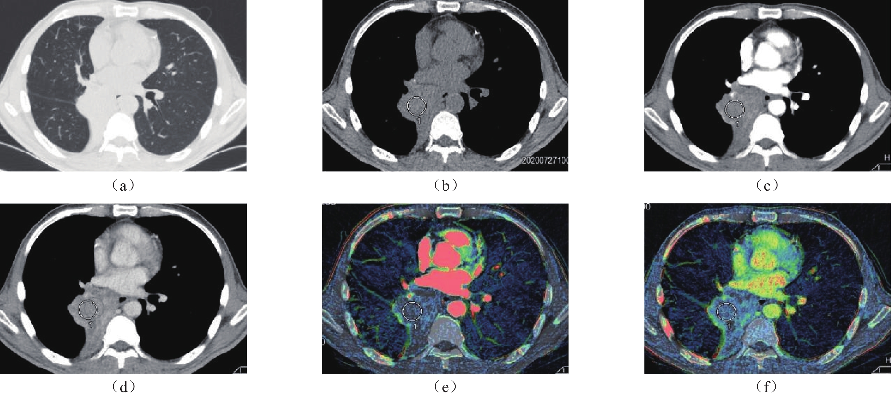
随后,3位研究者分别在动脉期、静脉期减影碘图上测量病灶的平均CT值。病灶肺减影CT图像的测量值分别记录为动脉期∆Husub、静脉期∆Husub,ROI绘制要求与普通增强CT一致(图1)。
1.4 统计学分析
采用SPSS 22.0软件进行统计学分析。正态分布计量资料采用平均值±标准差表示,非正态分布计量资料用中位数(四分位数间距)表示,计数资料以频数表示。使用组内相关系数(interclass correlation coefficient,ICC)分析3位研究者测量数据的一致性,如果一致性良好,则取3次测量的平均值进行进一步分析。采用McNemar检验和kappa检验分别评估动、静脉期∆Hu等级和减影CT碘图视觉评估灌注量等级的差异和一致性。采用配对t检验比较动、静脉期∆Hu和∆Husub之间的差异。P<0.05表明差异具有统计学意义。
2. 结果
本研究共纳入34例患者(37个病灶),其中男性23例,女性11例,患者平均年龄为(60.3±14.5)岁。在37个病灶中,28个位于肺内(右肺12个,左肺16个),9个位于纵隔。病灶的中位直径为6.7(5.6)cm。
3位观察者测量数据的一致性较好,ICC均>0.9(表1)。
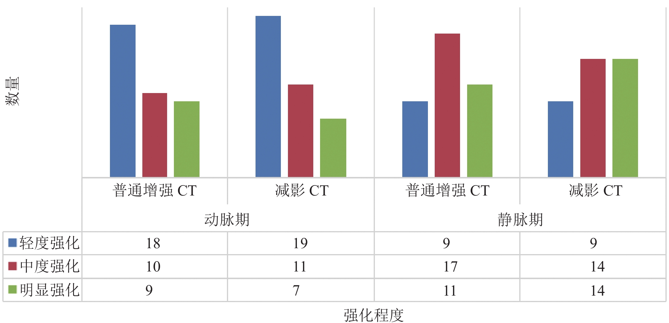
表 1 3位观察者测量减影CT与普通增强CT两期增强净增值的一致性Table 1. Consistency of net incremental value of enhanced CT with and without subtraction measured by three observers动脉期 静脉期 普通增强CT 减影CT 普通增强CT 减影CT ICC 0.985 0.986 0.947 0.971 图2所示为动、静脉期∆Hu等级和减影碘图视觉评估病灶灌注量等级的分布图。动脉期∆Hu等级和CT减影碘图视觉评估病灶灌注量等级之间无显著差异(P=0.223),且两者一致性较好(Kappa=0.870,P<0.001)。静脉期∆Hu等级和CT减影碘图视觉评估病灶灌注量等级之间也无显著差异(P=0.223),且具有中等一致性(Kappa=0.711,P<0.001)。
如表2所示,动、静脉期普通增强CT计算的∆Hu和CT肺减影碘图测量的∆Husub之间差异均无统计学意义(动脉期P=0.321,静脉期P=0.313)。肺内病灶和纵隔病灶动、静脉期普通增强CT计算的∆Hu和CT肺减影碘图测量的∆Husub之间的差异均无统计学意义(肺内病灶:动脉期P=0.595;静脉期P=0.793;纵隔病灶:动脉期P=0.265;静脉期P=0.306)。
表 2 动、静脉期普通增强CT计算的∆Hu和CT肺减影碘图测量的∆Husub的差异比较Table 2. Comparison of the difference between ∆Hu computed in common contrast-enhanced CT and ∆Husub measured in iodine maps generated from subtraction CT in the arterial and venous phases总(n=37) 肺内病灶(n=27) 纵隔病灶(n=10) 动脉期 静脉期 动脉期 静脉期 动脉期 静脉期 △Hu/HU 25.29±17.98 39.07±11.08 25.49±22.34 37.97±21.53 24.14±17.89 33.84±38.08 △Husub/HU 25.54±17.96 38.84±12.05 25.68±21.68 37.88±21.48 23.46±17.42 33.42±37.49 t 1.007 −1.024 −0.539 0.265 1.187 1.085 P 0.321 0.313 0.595 0.793 0.265 0.306 注:∆Hu为普通增强CT强化净增值,∆Husub为肺减影碘图灌注值,n代表病灶数量。 3. 讨论
CT肺减影技术是一种基于精确图像柔性配准算法的技术,其原理是从增强后CT图像中减去平扫CT图像,以获得有关碘分布的信息[11]。CT肺减影技术生成的彩图中碘分布浓度与病变/组织器官的碘摄取量成正比[12],且生成的碘分布图能以灌注伪彩图的形式直观显示出来,故可以显著提高人眼对图像密度差异的辨识能力[13],因此碘分布彩图可以反映两次扫描间病变/组织器官的密度差异,从而直观地显示出病变/组织器官的对比强化程度。
本研究的主要结果为,CT肺减影碘图视觉评估灌注量等级和普通增强CT强化程度无显著差异,且具有较好的一致性。肺减影CT测量的病灶灌注值(∆Husub)和普通增强CT测量的强化净增值(∆Hu)无明显差异。
CT减影技术在临床中的应用已被证实。Dagmar等[14]学者的一项研究结果显示,对比单纯肺动脉CT造影,联合CT减影碘图可以小幅度提高肺栓塞的诊断效能,诊断的特异性较单纯肺动脉CT造影略高(100% VS. 94%),且敏感性相似,这使假阳性诊断的可能性减小。Kunihiro等[15]学者使用CT减影冠状动脉造影评估冠状动脉重度钙化节段狭窄的效能高于普通冠状动脉CT造影(敏感性:94.1% VS. 88.2%;特异性:85.4% VS. 62.5%;准确性:89.0% VS. 73.2%)。这表明了CT减影技术为血管性病变的诊断提供了更多的额外信息,弥补了高密度病灶和高密度对比剂难以区分的缺点。也证明了肺减影CT在临床中的实用性和可行性。
然而,CT肺减影技术在胸部占位性病变中的应用研究较少。一项采用CT时间减影技术检测肺结节的研究结果显示,CT时间减影技术较肺CT平扫检出肺结节的敏感性提高了10%,且没有延长阅片时间[16]。这体现了对比于肺CT平扫,肺减影CT不但不增加阅片时间,还在描述组织密度差异方面具有优势。另一项关于减影CT和双能CT在肺结节(直径≥4 mm)强化中的对比研究,对30例患者存在的肺实性结节或肿块进行评估,结果显示每个研究者在减影CT中结节或肿块的检出率均比双能CT高,CT肺减影碘图中结节或肿块强化的可见性也更好[4]。这不但表明了减影CT有助于结节的检出,还表明了减影CT碘图可以更好的描述病灶的血流灌注情况。
在本研究中,减影CT碘图和普通增强CT的视觉评估和定量测量对胸部占位病变(无论病灶位于肺内或纵隔内)强化程度的评估均具有较好的一致性,且无显著统计学差异。这表明了CT肺减影碘图对胸部占位病变增强前、后灌注程度的描述能达到临床诊断需求,与普通增强CT无明显差异。尽管两者无统计学差异,但在视觉评估中普通CT和减影CT不同强化程度的病灶数量有所差异,普通CT动脉期明显强化病灶更多(9个VS. 7个),而静脉期相反,减影CT明显强化病灶更多(11个VS. 14个)。分析差异的原因,可能是因为减影CT可以更好的避免病灶区域伪影的影响,更直观的显示病灶强化程度[17];此外,减影技术采用高精度柔性配准,解剖配准精度高[18]。
本研究的不足之处:首先,本研究样本量较小,可能存在选择偏倚,未来应扩大样本量进一步研究;其次,没有计算采用减影CT和普通增强CT阅片的具体时长,但是通过阅片步骤和程序的简化,也可以间接反映减影CT可以在保证诊断需求的前提下,节省阅片及报告书写时间。
4. 结论
综上所述,肺减影CT和普通CT对胸部占位性病变强化程度的评估无显著差异,且肺减影CT可以在不增加扫描时长的前提下,简化阅片时对病灶进行测量和计算的步骤,有助于提高诊断效率。
-
表 1 3位观察者测量减影CT与普通增强CT两期增强净增值的一致性
Table 1 Consistency of net incremental value of enhanced CT with and without subtraction measured by three observers
动脉期 静脉期 普通增强CT 减影CT 普通增强CT 减影CT ICC 0.985 0.986 0.947 0.971 表 2 动、静脉期普通增强CT计算的∆Hu和CT肺减影碘图测量的∆Husub的差异比较
Table 2 Comparison of the difference between ∆Hu computed in common contrast-enhanced CT and ∆Husub measured in iodine maps generated from subtraction CT in the arterial and venous phases
总(n=37) 肺内病灶(n=27) 纵隔病灶(n=10) 动脉期 静脉期 动脉期 静脉期 动脉期 静脉期 △Hu/HU 25.29±17.98 39.07±11.08 25.49±22.34 37.97±21.53 24.14±17.89 33.84±38.08 △Husub/HU 25.54±17.96 38.84±12.05 25.68±21.68 37.88±21.48 23.46±17.42 33.42±37.49 t 1.007 −1.024 −0.539 0.265 1.187 1.085 P 0.321 0.313 0.595 0.793 0.265 0.306 注:∆Hu为普通增强CT强化净增值,∆Husub为肺减影碘图灌注值,n代表病灶数量。 -
[1] NIU Y, LIU W, XIAN L, et al. Thoracic splenosis presenting as pulmonary space-occupying lesion[J]. BMC Surgery, 2018, 18(1): 119. DOI: 10.1186/s12893-018-0461-9.
[2] SCORSETTI M, LEO F, TRAMA A, et al. Thymoma and thymic carcinomas[J]. Critical Reviews Oncology/Hematology, 2016, 99: 332-350. DOI: 10.1016/j.critrevonc.2016.01.012.
[3] 孙学术. 胸部增强CT检查在肺癌诊断及鉴别诊断中的应用研究进展[J]. 中国医疗器械信息, 2023, 29(2): 30-32. DOI: 10.3969/j.issn.1006-6586.2023.02.010. SUN X S. Advances in the application of enhanced chest CT in the diagnosis and differential diagnosis of lungcancer[J]. China Medical Device Information, 2023, 29(2): 30-32. DOI: 10.3969/j.issn.1006-6586.2023.02.010.
[4] GROB D, OOSTVEEN L J, JACOBS C, et al. Pulmonary nodule enhancement in subtraction CT and dual-energy CT: A comparison study[J]. European Journal of Radiology, 2021, 134: 109443. DOI: 10.1016/j.ejrad.2020.109443.
[5] JEONG Y J, LEE K S, JEONG S Y, et al. Solitary pulmonary nodule: Characterization with combined wash-in and washout features at dynamic multi-detector row CT[J]. Radiology, 2005, 237(2): 675-683. DOI: 10.1148/radiol.2372041549.
[6] 燕子, 蔡华崧, 胡曼诗, 等. 肝脏CT动态增强扫描非刚性对位减影技术的质量评价[J]. 中华普通外科学文献(电子版), 2019, 13(6): 459-462. DOI: 10.3877/cma.j.issn.1674-0793.2019.06.009. YAN Z, CAI H S, HU M S, et al. Quality evaluation of non-rigid subtraction technique in CT dynamic enhancement scanning of liver[J]. Chinese Archives of General Surgery (Electronic Edition) 2019, 13(6): 459-462. DOI: 10.3877/cma.j.issn.1674-0793.2019.06.009.
[7] 王晨思, 洪瑶, 方慧敏, 等. 基于自动管电流调制技术下不同螺距对颈部CTA辐射剂量及图像质量影响的研究[J]. CT理论与应用研究(中英文), 2024, 33(3): 309-315. DOI: 10.15953/j.ctta.2023.148. WANG C S, HONG Y, FANG H M, et al. Based on the effect of different pitches on neck CTA radiation dose and image quality under automatic tube current modulation technology[J]. CT Theory and Applications, 2024, 33(3): 309-315. DOI: 10.15953/j.ctta.2023.148.
[8] 王倩, 彭文廷, 王蓉, 等. CT特征及增强定量分析预测肺磨玻璃结节浸润性的诊断价值[J]. 医学影像学杂志, 2024, 34(4): 33-37,48. WANG Q, PENG W T, WANG R, et al. The value of CT features and quantitative analysis of contrast-enhanced CT scan in predicting the invasion of pulmonary ground-glass nodules[J]. Journal of Medical Imaging, 2024, 34(4): 33-37,48. (in Chinese).
[9] 陈超, 刘伟, 徐黎明, 等. 探究减影CT肺血管造影碘图在卵巢癌患者术前急性肺动脉栓塞诊断中的价值[J]. 肿瘤影像学, 2023, 32(2): 132-137. DOI: 10.19732/j.cnki.2096-6210.2023.02.005. CHEN C, LIU W, XU L M, et al. To explore the value of subtraction iodine mapping CT pulmonary angiography in the diagnosis of acutepulmonary embolism in preoperative patients with ovarian cancer[J]. Oncoradiology, 2023, 32(2): 132-137. DOI: 10.19732/j.cnki.2096-6210.2023.02.005.
[10] 王嘉瑞, 丁向明. 螺旋CT增强扫描在肺癌患者淋巴结转移诊断的应用价值[J]. 影像研究与医学应用, 2024, 8(3): 133-135. DOI: 10.3969/j.issn.2096-3807.2024.03.043. [11] GROB D, OOSTVEEN L J, PROKOP M, et al. Imaging of pulmonary perfusion using subtraction CT angiography is feasible in clinical practice[J]. European Radiology, 2019, 29(3): 1408-1414. DOI: 10.1007/s00330-018-5740-4.
[12] PELGRIM G J, VAN HAMERSVELT R W, WILLEMINK M J, et al. Accuracy of iodine quantification using dual energy CT in latest generation dual source and dual layer CT[J]. European Radiology, 2017, 27(9): 3904-3912. DOI: 10.1007/s00330-017-4752-9.
[13] 邢小明, 邓利华, 张春, 等. 640层CT肺减影成像联合肺动脉CTA诊断急性肺栓塞的初步探究[J]. 影像研究与医学应用, 2022, 6(5): 5-7. DOI: 10.3969/j.issn.2096-3807.2022.05.002. XING X M, DENG L H, ZHANG C, et al. Preliminary study on 640-slice CT lung subtraction imaging combined with pulmonary CTA in the diagnosis of acute pulmonaryembolism[J]. Journal of Imaging Research and Medical Applications, 2022, 6(5): 5-7. DOI: 10.3969/j.issn.2096-3807.2022.05.002.
[14] GROB D, SMIT E, PRINCE J, et al. Iodine maps from subtraction CT or dual-energy CT to detect pulmonary emboli with CT angiography: A multiple-observer study[J]. Radiology, 2019, 292(1): 197-205. DOI: 10.1148/radiol.2019182666.
[15] YOSHIOKA K, TANAKA R, MURANAKA K, et al. Subtraction coronary CT angiography using second-generation 320-detector row CT[J]. The International Journal of Cardiovascular Imaging, 2015, 31(S1): 51-58. DOI: 10.1007/s10554-015-0630-1.
[16] AOKI T, MURAKAMI S, KIM H, et al. Temporal subtraction method for lung nodule detection on successive thoracic CT soft-copy images[J]. Radiology, 2014, 271(1): 255-261. DOI: 10.1148/radiol.13130460.
[17] 王之龙, 朱旭, 高顺禹, 等. CT减影法评价肝细胞癌TACE后残余肿瘤活性的研究[J]. 介入放射学杂志, 2017, 26(1): 64-68. DOI: 10.3969/j.issn.1008-794X.2017.01.014. WANG Z L, ZHU X, GAO S Y, et al. Application of CT subtraction technique in evaluating residual hepatocelluar carcinoma activity after transcatheter arterial chemoembolization[J]. Journal of Interventional Radiology, 2017, 26(1): 64-68. DOI: 10.3969/j.issn.1008-794X.2017.01.014.
[18] 朱乐, 雷子乔, 余建明, 等. 320排CT肺血管减影成像对急性肺栓塞诊断的初步临床研究[J]. 临床放射学杂志, 2017, 36(5): 728-732. ZHU L, LEI Z Q, YU J M, et al. Preliminary Study on 320-Row CT Digital Subtraction Pulmonary Angiography in Acute Pulmonary Embolism[J]. Journal of Clinical Radiology, 2017, 36(5): 728-732. (in Chinese).
-
期刊类型引用(0)
其他类型引用(1)


 下载:
下载:


