Application Value of Variable Exposure Conditions in One-stop Cerebral Computed Tomography Imaging
-
摘要:
目的:通过比较固定曝光条件和可变曝光条件在一站式颅脑CT扫描中的影像质量和辐射剂量,探讨可变曝光条件在一站式颅脑CT扫描中的应用价值。方法:前瞻性选取我院2024年3至2024年5月间,因脑血管疾病需行头颅一站式CT扫描的患者100例,随机分为A组和B组,每组各50例。所有患者均从对比剂注射后5 s开始第1期扫描,至65 s结束,共进行18期扫描。A组患者所有扫描期相均使用相同的曝光条件,即管电压100 kV,管电流量100 mAs。B组根据各期相影像数据的诊断目的不同,使用不同的管电流量进行扫描。使用CT后处理工作站,测量平扫期大脑半球灰质、白质及颅外空气的CT值和噪声,计算图像的对比噪声比(CNR);测量CTA图像的颅内动脉、脑实质区的CT值和噪声,计算CNR;测量CTP图像的CBV、CBF、MTT、TTP及Tmax>6 s的脑组织体积等灌注参数,并对各期图像质量进行主观评价。使用Shapiro-Wilk检验客观指标的正态性,两组间客观指标比较采用独立样本t检验进行。采用Wilcoxon符号秩检验比较主观评分结果。结果:B组平扫图像的CNRp和主观评分优于A组。CTA图像噪声小于A组,CNRa及主观评分均高于A组;B组CTP各灌注参数与A组无差异;B组患者的DLP较A组增加约0.65%。结论:与固定曝光条件组相比,可变曝光条件一站式颅脑CT的平扫和CTA图像质量均明显提高,各灌注参数无明显差异,总体辐射剂量无明显增加的情况下,提供比固定曝光条件更好的图像质量。
Abstract:Objective: To compare the imaging quality and radiation dose under fixed and variable exposure conditions and to determine the value of variable exposure conditions in a one-stop cranial computed tomography (CT). Methods: One hundred patients who required one-stop head CT scanning because of cerebrovascular disease at our hospital between March and May 2024 were prospectively selected and randomly divided into groups A and B, with 50 patients in each group. All patients underwent the first phase of scanning at 5~65s after contrast injection, and a total of 18 scanning phases were performed. In group A, all patients underwent the same exposure conditions for all scan phases, with a tube voltage of 100kV and tube current of 100mAs. In group B, the patients were scanned with different tube currents according to the different diagnostic purposes of each phase of the image data. The CT values and noise of the gray matter, white matter, and extra-cranial air in the cerebral hemisphere during normal scans were measured using CT post-processing workstation, and the contrast noise ratio (CNR) of the images was calculated. The CT values and noise in the intracranial artery and parenchyma were measured using computed tomography angiography (CTA) images, and the CNR was calculated. Perfusion parameters such as cerebral blood volume (CBV), cerebral blood flow (CBF), mean transit time (MTT), and time to peak (TTP) in the computer-to-plate-performed image were measured, and a five-point scale was used to subjectively evaluate the image quality at each stage. The Shapiro–Wilk test was performed to test the normality of objective indicators, and an independent sample t-test was used to compare objective indicators between the two groups. The Wilcoxon signed-rank test was used to compare subjective scores. Results: The CNRp and subjective scores of the plain scan images in group B were better than those in group A. The CTA image noise in group B was lower than that in group A, and the CNRa and subjective score in group B was higher than that in group A. However, the perfusion parameters of CTP in group B did not differ significantly from those in group A. The dose length product (DLP) in group B was increased by approximately 0.65% compared with that in group A. Conclusion: Compared with the fixed exposure condition group, the image quality of the one-stop cranial CT under variable exposure conditions was significantly improved. No significant differences were observed in any perfusion parameter, and the overall radiation dose was not significantly increased, providing better image quality than that under fixed exposure conditions.
-
脑血管疾病中具有高发病率、高致残率和高死亡率,是我国居民的第1位死亡原因[1-2]。随着宽体探测器CT的发展,通过一次对比剂注射,一次扫描即可完成颅脑CT平扫、CTA和CT灌注(CT perfusion,CTP)的一站式CT扫描成为急性脑血管病诊治的重要影像检查方法。然而CTP需进行多期扫描,扫描野内有晶状体等辐射敏感器官,多期扫描带来的辐射剂量问题不容忽视。如何在不增加或降低辐射剂量的前提下,提高一站式CT扫描的图像质量,受到越来越多的关注[3-5]。
我们通过对比研究不同曝光条件下一站式颅脑CT扫描的图像质量和辐射剂量,探讨可变曝光条件在一站式颅脑CT扫描中的应用价值。
1. 资料与方法
1.1 一般资料
前瞻性收集我院2024年3至2024年5月间因脑血管疾病需行一站式颅脑CT扫描的患者100例。采用区组长度为2,区组内顺序固定的区组随机分组法将100例患者随机分为A组和B组,每组50例。A组为固定曝光条件组,B组为可变曝光条件组。
病例纳入标准:临床怀疑脑血管疾病需行一站式CT扫描的患者。排除标准:①躁动无法配合检查的患者;②双侧颈内动脉或大脑中动脉均存在明显狭窄的患者;③有碘对比剂使用禁忌证的患者。
所有患者检查前均签署增强检查知情同意书。本研究经过南京大学医学院附属鼓楼医院医学伦理委员会批准(伦理号为2023-341-01)。
1.2 仪器设备方法
使用联影公司UIH 960 CT,欧力奇公司CT高压注射器。患者去除头部金属异物,仰卧位,固定头部,双上肢自然置于身体两侧。根据头颅Z轴方向的长度,准直宽度使用280×0.5 mm或320×0.5 mm,单圈轴扫完成全颅成像。
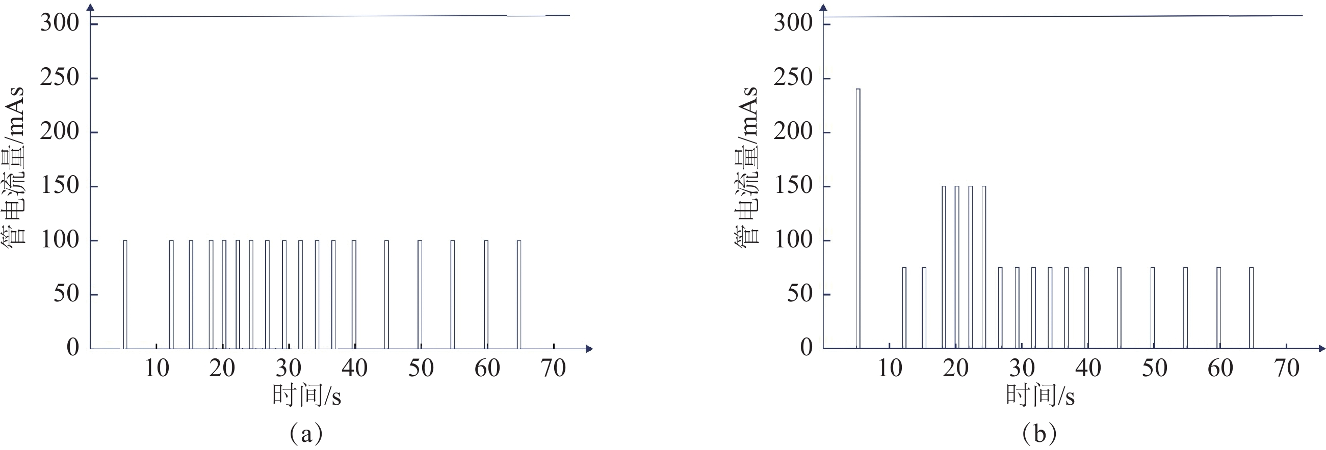
扫描参数。A组所有期相均使用相同的曝光条件,管电压100 kV,管电流量100 mAs,旋转时间0.5 s/r。从对比剂注射后5 s开始第1期扫描,至65 s结束,共进行18期扫描。第1期(平扫期)起始时间5.0 s,至5.5 s结束。第2~3期(动脉早期)起始时间12.0 s,结束时间15.5 s,间隔时间3.0 s;第4~7期(CTA峰值期)起始时间18.0 s,结束时间24.5 s,间隔时间2.0 s;第8~12期(动脉晚期)起始时间26.5 s,结束时间37.0 s,间隔时间2.5 s;第13~18期(平衡期)起始时间39.5 s,结束时间65.0 s,间隔时间5.0 s(图1(a))。
B组管电压和旋转时间与A组相同,并根据各期相影像数据的诊断目的不同,使用不同的管电流量进行扫描,平扫期(第1期)管电流量240 mAs,CTA峰值期(第4~7期)150 mAs,其余期相均为75 mAs(图1(b))。两组其余扫描参数均一致。
图像重建参数。滤过算法为H-Soft-B,迭代重建等级5级,窗宽80,窗位40。厚层图像层厚、层间隔均为5 mm,薄层图像层厚、层间隔均为1 mm,矩阵512×512。对比剂为碘佛醇(320 mgI/mL),注射速率5.0 mL/s,总量50 mL,结束后以3.0 mL/s速率跟注生理盐水30 mL。
1.3 图像处理和评价
将图像传至联影CT图像处理工作站(uWS-CT R004)进行分析。
主观评价均由两名从事放射诊断工作5年以上的医师共同进行评价,两人评分不一致时,则由第3名高年资医师确定最终评分结果,评分≥3分为满足临床诊断要求,评分≤2分无法满足诊断要求。
1.3.1 颅脑平扫图像质量评价
CTP第1期扫描于对比剂注射后5 s时进行,此时对比剂尚未进入体循环到达颅脑。选择该期图像作为头颅平扫图像进行质量评价。
客观评价。选取基底节层面,ROI大小约为10~20 mm2,避开伪影及病变区域,分别测量健侧大脑半球灰质、白质以及颅外空气的CT值及其标准差(standard deviation,SD),SD值作为噪声值。以灰、白质噪声的平均值作为平扫图像噪声(SDp),并以颅外空气的噪声作为图像的背景噪声(SD1)[6]。
计算平扫图像的对比噪声比(contrast to noise ratio,CNR),CNRp=(灰质CT值−白质CT值)/背景SD1。
主观评价。由两名放射诊断医师共同对平扫图像进行评价,评价内容包括灰白质对比度、骨脑交界处硬化伪影干扰情况、梗死灶及出血灶的显示、主观噪声等。
上述评价指标按照5分法评定。评分标准[7]:5分,脑灰白质对比良好,结构显示清晰,图像噪声较小,无明显伪影;4分,脑灰白质对比良好,结构显示清晰,图像噪声适中,可有轻微伪影但不影响诊断;3分,脑灰白质对比尚可,结构显示较清晰,图像噪声适中,有少许伪影,基本满足诊断要求;2分,脑灰白质对比稍差,部分结构显示不清,图像噪声稍大,部分层面有少许伪影,诊断信心不足;1分,脑灰白质对比较差,部分结构显示不清,图像噪声明显,部分层面有严重伪影,无法满足诊断要求。
1.3.2 CTA图像处理和评价
将1 mm的薄层图像导入血管处理软件,选择大脑中动脉或大脑前动脉放置ROI,软件自动生成动脉血管的时间密度曲线(time density curve,TDC),选择CT值到达峰值前的一期图像进行CTA三维成像。
客观评价。测量颅内动脉的CT值及其噪声(SDa),首选右侧颈内动脉进行测量和评估。若右颈内动脉有明显狭窄或闭塞,则选择左颈内动脉进行测量,若双侧颈内动脉均有明显病变,则选择椎动脉末端进行测量,ROI直径约为血管直径的1/2。测量同侧脑实质区CT值及其噪声(SD2),ROI大小约为10~20 mm2。计算CTA图像的CNR,CNRa=(动脉CT值−脑实质CT值)/脑实质噪声SD2。
主观评价。由上述两名诊断医师共同对CTA图像进行主观评价,评价内容包括:动脉血管和周围结构的对比度、管壁锐利程度、侧支循环显示情况、主观噪声等,上述评价指标均按照5分法评定。评分标准[8]:5分,颈部及头部动脉血管内对比剂充盈良好,血管连续,轮廓光滑,血管壁边缘锐利,无伪影,图像噪较小;4分,颈部及头部动脉血管对比剂充盈良好,血管连续,轮廓清楚,管壁轻微模糊,或有轻微伪影,可用于诊断;3分,颈部及头部动脉血管充盈尚可,血管连续性尚可,管壁稍毛糙,或有轻度伪影,图像噪声稍大,基本满足诊断要求;2分,颈部及头部动脉血管充盈差或连续性欠佳,管壁毛糙,有较多伪影,图像噪声较大,诊断受限;1分,颈部及头部动脉血管不能识别,连续性中断,伪影重,图像噪声较大,无法诊断。
1.3.3 CTP图像处理和评价
两名诊断医生采用基于去卷积算法的脑灌注分析软件,对5 mm层厚的CTP图像进行数据分析。
客观评价。通过软件自动识别输入动脉和输出静脉,并由人工进行核对。参考CTA图像评价方法,选择合适的动脉作为输入动脉,避免选择有明显病变的血管,选择上矢状窦作为输出静脉,并绘制动、静脉的TDC曲线。在正常侧的脑灌注伪彩图上,选择基底节层面的颞叶白质及尾状核头区分别放置面积为约20 mm2的ROI,计算脑血流量(cerebral blood flow,CBF)脑血容量(cerebralblood volume,CBV)、平均通过时间(mean transit time,MTT)、达峰时间(time to peak,TTP)等灌注参数,以及残余组织达峰时间(time to maximum of the residual function,Tmax)>6 s时的脑组织体积。
主观评价。由上述两名诊断医师共同对CTP图像进行主观评价。根据Li等[9]相关图像质量标准,将灌注伪彩图像进行评分:5分,灌注图像质量好,明显区分灰白质,无缺损区,完全可以诊断;4分,灌注图像质量较好,可以区分灰白质,无缺损区,可以诊断;3分,灌注图像质量一般,仍能区分灰白质,有较少缺损区,可以诊断;2分,灌注图像质量较差,灰白质难以区分,缺损区较多,基本可以诊断;1分,灌注图像质量差,缺损区很多,无法诊断。
1.4 辐射剂量
记录A组和B组患者的扫描长度、CT容积计量指数(CT dose index volume,CTDIvol)和剂量长度乘积(dose length product,DLP)。
1.5 统计学分析
采用SPSS统计软件进行统计学分析。采用Shapiro-Wilk检验计量资料的正态性,以均值±标准差
$( \bar x\pm s ) $ 表示符合正态分布的数据,两组间比较采用独立样本t检验。对两组图像的灰质CT值、白质CT值、图像噪声、背景噪声和CNR采用独立样本t检验;图像质量主观评分以中位数(上、下四分位数)M(Q1,Q3)表示,采用Kappa分析比较两名医师主观评分的一致性,Kappa值<0.4为一致性较差,0.4≤Kappa值<0.75为一致性中等,Kappa值≥0.75为一致性好。采用Wilcoxon符号秩检验比较主观评分结果。P<0.05认为差异具有统计学意义。
2. 结果
2.1 两组患者一般资料及辐射剂量比较
A组和B组患者的性别、年龄、身高、体质量、扫描长度均无统计学差异。
B组患者较A组患者的CTDIvol增加了0.5%,DLP增加了0.9%,差异具有统计学意义(表1)。
表 1 两组患者一般资料及辐射剂量比较Table 1. General information and radiation dose comparison of the two patient groups项目 组别 统计检验 A组 B组 统计值 P 例数 50 50 - - 性别 男 28 27 0.400a 0.841 女 22 23 评价年龄/岁 62.8±13.5 60.3±12.1 0.997 0.321 身高/m 1.66±0.07 1.65±0.06 0.899 0.371 体质量/kg 68.5±9.1 69.8±8.9 0.773 0.441 扫描长度/cm 140.40±2.83 140.80±3.96 − 0.581 0.562 CTDIvol/mGy 152.20±0.15 153.09±0.412 −14.232 <0.001 DLP/(mGy·cm) 2136.74 ±39.822156.52 ±57.05−2.010 0.047 注:CTDI为CT剂量指数;DLP为剂量长度乘积,a为$\chi^2 $值,其他为t值。 2.2 平扫图像质量分析
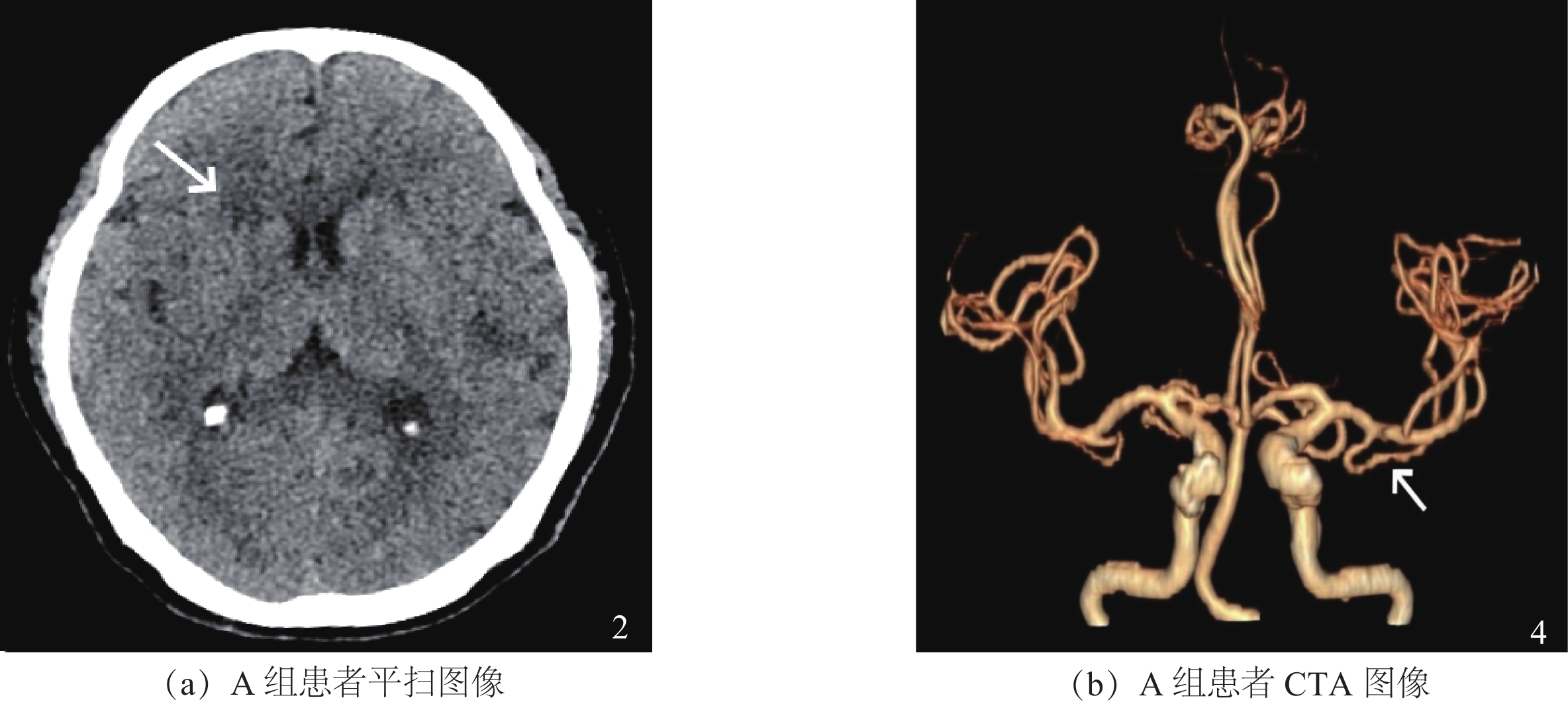
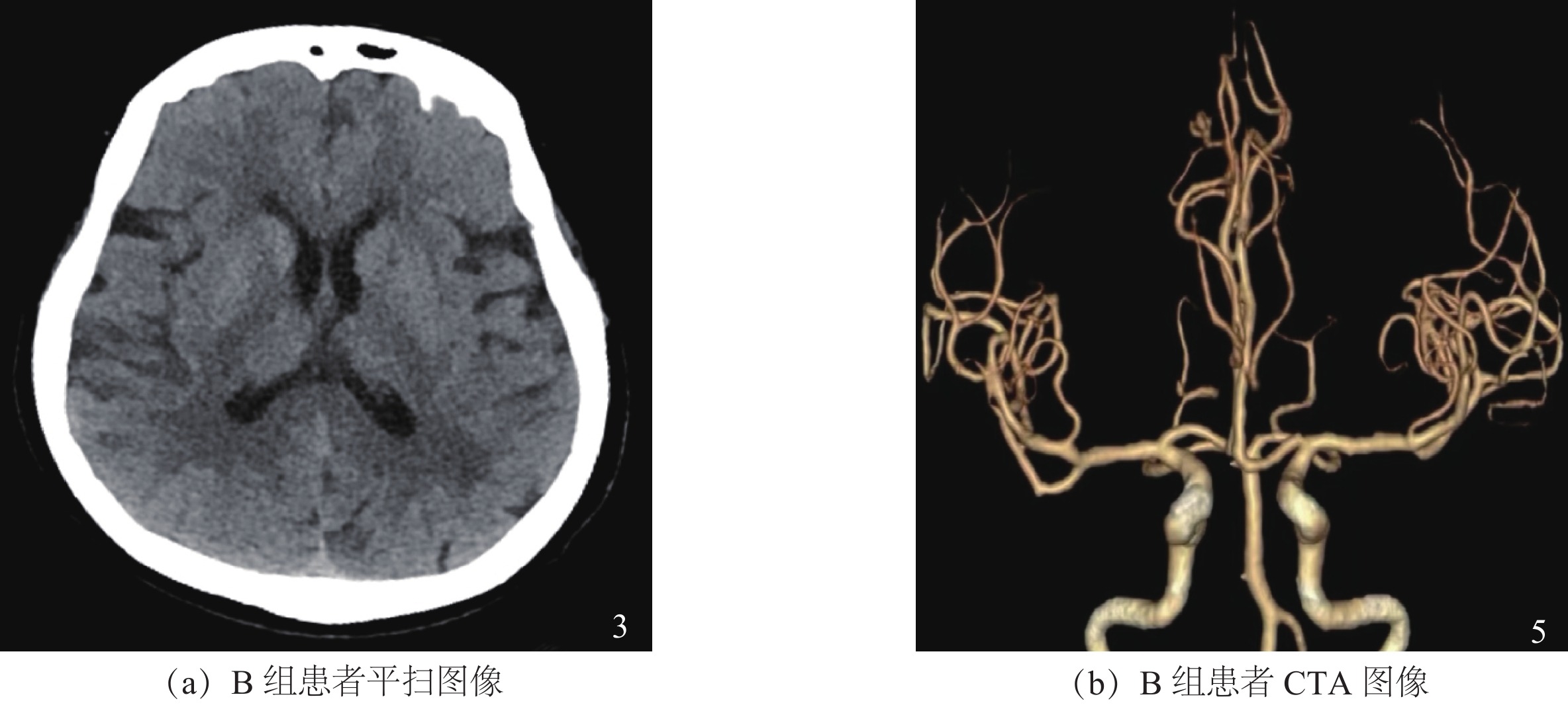
A组和B组平扫图像脑灰质、白质的CT值均无统计学差异。B组图像噪声和背景噪声较A组分别下降了59.6%和46.7%,CNRp比A组提高100.5%,图像质量评分大于A组(图2(a)和图3(a)),差异均具有统计学意义(表2)。
表 2 两组患者平扫图像质量比较Table 2. Comparison of plain scan image quality between the two patient groups项目 组别 统计检验 A组 B组 统计值 P 灰质CT值 36.41±1.51 36.15±1.26 0.896 0.370 白质CT值 27.14±1.69 26.85±1.54 0.831 0.408 图像噪声SDp 4.75±0.78 1.92±0.22 8.537 0.000 背景噪声SD1 3.19±0.55 1.70±0.28 16.826 0.000 CNRp 10.33±2.11 20.72±3.53 −8.473 0.000 评分 3(3,3) 5(4,5) −8.681a 0.000 注:a为Z值,其余统计值均为t值。 两名医师的图像质量主观评分具有较高的一致性,Kappa值分别为0.77和0.81。
2.3 CTA图像质量分析
A组和B组CTA图像颅内动脉、脑实质的CT值均无统计学差异。B组动脉噪声和图像噪声较A组分别下降了23.2%和29.6%,CNRp比A组提高48.3%,图像质量评分大于A组(图2(b)和图3(b)),差异均具有统计学意义(表3)。
表 3 两组患者CTA图像质量比较Table 3. Comparison of CTA image quality between the two patient groups项目 组别 统计检验 A组 B组 统计值 P 颅内动脉CT值 391.64±64.57 409.61±72.56 1.113 0.269 动脉噪声SDa 7.00±0.73 5.37±0.65 −6.963 0.000 脑实质CT值 36.53±1.61 36.32±1.66 −0.546 0.586 图像噪声SD2 9.13±1.44 6.42±0.46 −7.414 0.000 CNRa 39.53±8.23 58.63±12.93 8.021 0.000 评分 4(3,4) 5(5,5) 5.745a 0.000 注:a为Z值,其余统计值均为t值。 两名医师的图像质量主观评分具有较高的一致性,Kappa值分别为0.88和0.83。
2.4 CTP图像质量分析
A组和B组CTP图像尾状核头及颞叶白质的CBV、CBF、MTT、TTP及Tmax>6 s的脑组织体积均无统计学差异(表4)。
表 4 两组患者CTP结果和图像质量比较Table 4. Comparison of CTP results and image quality between the two patient groups项目 组别 统计检验 A组 B组 统计值 P 尾状核头 CBV 3.54±0.96 3.63±1.16 − 0.010 0.992 CBF 99.22±40.91 107.25±53.37 0.283 0.777 MTT 3.94±1.53 3.79±1.58 − 0.430 0.668 TTP 19.13±2.79 18.31±2.41 −1.391 0.168 颞叶白质 CBV 1.98±0.72 2.00±0.79 − 0.152 0.88 CBF 45.03±18.42 42.76±23.26 −1.131 0.258 MTT 5.55±2.30 5.59±2.07 0.47 0.639 TTP 20.58±2.81 20.39±2.55 − 0.316 0.753 评分 4(4,5) 4(4,5) −1.076a 0.282 Tmax>6 s的脑组织体积/mL 74.19±126.79 34.56±78.94 1.876 0.064 注:a为Z值,其余统计值均为t值。 两名医师的图像质量主观评分具有较高的一致性,Kappa值分别为0.87和0.80。
3. 讨论
在一站式颅脑CT扫描中,CT平扫主要用于出血性脑血管病和缺血性脑血管病的鉴别、梗死灶的评估以及ASPECT评分等[10-12]。CTA主要用于判断病因,明确责任血管,评估侧支循环情况,能够更全面地显示血管形态。相较于DSA对血管产生刺激作用而引起多种检查相关不良反应,CTA更加安全可靠[13]。CTP主要是利用多种灌注参数的定量分析,评估脑组织微循环的灌注状态,从而精确评估脑缺血区的范围大小及其严重程度。临床工作中,CTA与CTP两者联合应用具有更准确的评估价值,能够弥补血流灌注随时间而变化的不足,为卒中等脑血管病患者治疗策略的选择,提供更充分的影像学支持[14-16]。
一站式颅脑CT成像不同成像阶段的检查目的不同,对图像质量要求亦不相同。相对于颅脑平扫和CTA而言,CTP对图像的噪声、SNR等客观指标要求相对较低。既往研究显示,可通过优化采集流程,减少扫描期相、降低管电流量及管电压等方法来降低CTP的辐射剂量[17]。在固定曝光条件的一站式扫描中,所有期相使用相同的曝光条件,为了控制整个扫描的辐射剂量在可接受的范围,往往使用较低的曝光条件[18]。通过降低管电流量尽管可以降低辐射剂量,但过低的管电流量会造成图像伪影的产生和噪声的增加[19],从而导致平扫和CTA的图像质量较差,甚至影响诊断。
因此本研究使用的可变曝光条件的一站式扫描,每一个扫描阶段的管电流量都可以独立设置。对于图像质量要求较高的平扫和CTA期,使用较高的曝光条件,与固定曝光条件组相比,平扫和CTA期的图像质量都明显提高,提升医生的诊断信心。
本研究在对CTA图像进行分析时,我们并未选择动脉CT值到达峰值时的图像进行CTA图像评估,这是因为随着动脉CT值的不断升高,颅内静脉逐渐显影,当动脉CT值到达峰值时,静脉内已有大量对比剂,存在静脉污染现象,影响CTA成像效果。因此我们选择动脉CT值到达峰值前的一期图像进行CTA评估,以获得更高质量的CTA图像。而CTP图像主要用于计算血流灌注参数,对图像噪声的宽容度较高,可以使用较低的曝光条件,我们研究的结果显示,固定曝光条件及可变曝光条件两组CTP的灌注参数均无统计学差异,满足诊断要求。
本研究存在的局限性:①本研究B组对CTA期(第4~7期)采用较高的管电流量进行扫描,是否可以更加精准判断CTA期,减少使用高剂量扫描的期数,进一步降低辐射剂量,有待于进一步研究;②本研究共进行18期扫描,与他人关于降低颅脑CTP辐射剂量的研究相比,仍有进一步减少采集次数,降低辐射剂量的空间。
综上所述,相较于固定曝光条件,可变曝光条件的一站式颅脑CT,在没有明显增加辐射剂量的前提下,显著提高了平扫和CTA的图像质量,同时保证灌注图像质量和结果无差异,为临床准确评估病情,制定个体化治疗方案提供影像学依据。
-
表 1 两组患者一般资料及辐射剂量比较
Table 1 General information and radiation dose comparison of the two patient groups
项目 组别 统计检验 A组 B组 统计值 P 例数 50 50 - - 性别 男 28 27 0.400a 0.841 女 22 23 评价年龄/岁 62.8±13.5 60.3±12.1 0.997 0.321 身高/m 1.66±0.07 1.65±0.06 0.899 0.371 体质量/kg 68.5±9.1 69.8±8.9 0.773 0.441 扫描长度/cm 140.40±2.83 140.80±3.96 − 0.581 0.562 CTDIvol/mGy 152.20±0.15 153.09±0.412 −14.232 <0.001 DLP/(mGy·cm) 2136.74 ±39.822156.52 ±57.05−2.010 0.047 注:CTDI为CT剂量指数;DLP为剂量长度乘积,a为$\chi^2 $值,其他为t值。 表 2 两组患者平扫图像质量比较
Table 2 Comparison of plain scan image quality between the two patient groups
项目 组别 统计检验 A组 B组 统计值 P 灰质CT值 36.41±1.51 36.15±1.26 0.896 0.370 白质CT值 27.14±1.69 26.85±1.54 0.831 0.408 图像噪声SDp 4.75±0.78 1.92±0.22 8.537 0.000 背景噪声SD1 3.19±0.55 1.70±0.28 16.826 0.000 CNRp 10.33±2.11 20.72±3.53 −8.473 0.000 评分 3(3,3) 5(4,5) −8.681a 0.000 注:a为Z值,其余统计值均为t值。 表 3 两组患者CTA图像质量比较
Table 3 Comparison of CTA image quality between the two patient groups
项目 组别 统计检验 A组 B组 统计值 P 颅内动脉CT值 391.64±64.57 409.61±72.56 1.113 0.269 动脉噪声SDa 7.00±0.73 5.37±0.65 −6.963 0.000 脑实质CT值 36.53±1.61 36.32±1.66 −0.546 0.586 图像噪声SD2 9.13±1.44 6.42±0.46 −7.414 0.000 CNRa 39.53±8.23 58.63±12.93 8.021 0.000 评分 4(3,4) 5(5,5) 5.745a 0.000 注:a为Z值,其余统计值均为t值。 表 4 两组患者CTP结果和图像质量比较
Table 4 Comparison of CTP results and image quality between the two patient groups
项目 组别 统计检验 A组 B组 统计值 P 尾状核头 CBV 3.54±0.96 3.63±1.16 − 0.010 0.992 CBF 99.22±40.91 107.25±53.37 0.283 0.777 MTT 3.94±1.53 3.79±1.58 − 0.430 0.668 TTP 19.13±2.79 18.31±2.41 −1.391 0.168 颞叶白质 CBV 1.98±0.72 2.00±0.79 − 0.152 0.88 CBF 45.03±18.42 42.76±23.26 −1.131 0.258 MTT 5.55±2.30 5.59±2.07 0.47 0.639 TTP 20.58±2.81 20.39±2.55 − 0.316 0.753 评分 4(4,5) 4(4,5) −1.076a 0.282 Tmax>6 s的脑组织体积/mL 74.19±126.79 34.56±78.94 1.876 0.064 注:a为Z值,其余统计值均为t值。 -
[1] 彭斌, 刘鸣, 崔丽英, 等. 中国急性缺血性脑卒中诊治指南2018[J]. 中华神经科杂志, 2018, 51(9): 666-682. DOI: 10.3760/cma.j.issn.1006-7876.2018.09.004. PENG B, LIU M, Cui L Y, et al. interpretation of the Chinese guidelines for diagnosis and treatment of acute ischemic stroke 2018[J]. Chinese Journal of Neurology, 2018: 657-659. DOI:10.3760/cma.j.issn.1006-7876.2018.09.004. (in Chinese).
[2] 中华医学会影像技术分会. 急性脑卒中多层螺旋CT检查技术专家共识[J]. 中华放射学杂志, 2020, 54(9): 839-845. DOI: 10.3760/cma.j.cn112149-20191226-01008. Chinese Society of Imaging Technology Chinese Medical Association. Expert consensus on multi-slice spiral CT examination for acute stroke[J]. 2020, 54(9): 839-845. DOI:10.3760/cma.j.cn112149-20191226-01008. (in Chinese).
[3] 于蒙蒙, 任昕晨, 邓雯雯, 等. 全脑CT灌注联合头颈部CTA“一站式”检查在前循环急性缺血性脑卒中的诊断价值[J]. 中国CT和MRI杂志, 2023, 21(7): 8-11. DOI: 10.3969/j.issn.1672-5131.2023.07.003. YU M M, REN X C, DENG W W, et al. The value of whole brain CT perfusion imaging combined with head and neck CTA one-stop examination in the diagnosis of anterior circulation acute ischemic stroke[J] Chinese Journal of CT and MRI, 2023, 21(7): 8-11. DOI:10.3969/j.issn.1672-5131.2023.07.003. (in Chinese).
[4] BIVARD A, PARSONS M. Tissue is more important than time: Insights into acute ischemic stroke from modern brain imaging[J]. Current Opinion in Neurology, 2018, 31(1): 23-27. DOI: 10.1097/WCO.0000000000000520.
[5] 蔡培, 徐凯, 牛磊, 等. 双低技术在iCT全脑灌注成像中的应用研究[J]. 医学影像学杂志, 2019, 29(10): 1656-1660. DOI: CNKI:SUN:XYXZ.0.2019-10-009. CAI P, XU K, NIU L, et al. Case study about the application of the double low techniques on the whole brain iCT perfusion imaging[J]. Journal of Medical Imaging, 2019, 29(10): 1656-1660. DOI:CNKI:SUN:XYXZ.0.2019-10-009. (in Chinese).
[6] 李文, 张志伟, 左子钰, 等. 双层探测器光谱CT虚拟单能量CTA技术对脑血管成像的价值[J]. CT理论与应用研究(中英文), 2024, 33(6): 669-675. DOI: 10.15953/j.ctta.2024.074. LI W, ZHANG Z W, ZUO Z Y, et al. The value of virtual monoenergetic computed tomography angiography with dual-layer detector spectral computed tomography for imaging cerebral vessels[J]. CT Theory and Applications, 2024, 33(6): 669-675. DOI: 10.15953/j.ctta.2024.074. (in Chinese).
[7] OZDOBA C, SLOTBOOM J, SCHROTH G, et al. Dose reduction in standard head CT: First results from a new scanner using iterative reconstruction and a new detector type in comparison with two previous generations of multi-slice CT[J]. Clinical Neuroradiology, 2014, 24(1): 23-28. DOI: 10.1007/s00062-013-0263-5.
[8] 杨尚文, 邵明冉, 杨献峰, 等. 三低"技术联合全模型迭代重建算法在头颈部CT血管成像中的可行性研究[J]. 中华放射医学与防护杂志, 2017, 37(1): 62-67. DOI: 10.3760/cma.j.issn.0254-5098.2017.01.012. YANG S W, SHAO M R, YANG X F, et al. A feasibility study on " Tri-Low" technology in combination with iterative model reconstruction (IMR) algorithm in CT angiography (CTA) of the head-and-neck vessels[J]. Chinese Journal of Radiological Medicine and Protection, 2017, 37(1): 62-67. DOI: 10.3760/cma.j.issn.0254-5098.2017.01.012. (in Chinese).
[9] LI Z L, LI H, ZHANG K, et al. Improvement of image quality and radiation dose of CT perfusion of the brain by means of low-tube voltage (70kV)[J]. European Radiology, 2014, 24(8): 1906-1913. DOI: 10.1007/s00330-014-3247-1.
[10] FURIE K L, JAYARAMAN M V. 2018 guidelines for the early management of patients with acute ischemic stroke[J]. Stroke, 2018, 49(3): 509-510. DOI: 10.1161/STROKEAHA.118.020176.
[11] 黄晓颖, 暴云锋, 李霞敏, 等. 人工智能在基于颅脑 CT灌注数据血管后处理的应用[J]. 中华放射学杂志, 2021, 55(8): 817-822. DOI: 10.3760/cma.j.cn112149-20200914-01087. HUANG X Y, BAO Y F, LI X M, et al. Application of artificial intelligence in vascular reconstruction based on cerebral CT perfusion data[J]. Chinese Journal of Radiology, 2021, 55(8): 817-822. DOI: 10.3760/cma.j.cn112149-20200914-01087. (in Chinese).
[12] WARNER J J, HARRINGTON R A, SACCO R L, et al. Guidelines for the early management of patients with acute ischemic stroke: 2019 update to the 2018 guidelines for the early management of acute ischemic stroke[J]. Stroke, 2019, 50(12): 3331-3332. DOI: 10.1161/STR.0000000000000211. (in Chinese).
[13] 刘青, 李伟粟, 王娇娇, 等. 全脑CT灌注成像在侧枝循环评估中的辐射剂量和临床应用价值[J]. 中华放射医学与防护杂志, 2024, 44(1): 47-52. DOI: 10.3760/cma.j.cn112271-20230609-00187. LIU Q, LI W L, WANG J J, et al. Radiation dose and clinical value of whole-brain CT perfusion imaging in the assessment of collateral circulation[J]. Chinese Journal of Radiological Medicine and Protection, 2024, 44(1): 47-52. DOI: 10.3760/cma.j.cn112271-20230609-00187. (in Chinese).
[14] FANG X K, NI Q Q, SCHOEPF U J, et al. Image quality, radiation dose and diagnostic accuracy of 70kVp whole brain volumetric CT perfusion imaging: A preliminary study[J]. European Radiology, 2016, 26(11): 4184-4193. DOI: 10.1007/s00330-016-4225-6.
[15] 逯瑶, 李玲, 曹若瑶, 等. 四维CT血管造影-CT灌注成像评价烟雾病及烟雾综合征侧支循环及其与脑血流动力学关系的研究[J]. 中华放射学杂志, 2023, 57(3): 252-258. DOI: 10.3760/cma.j.cn112149-20220401-00296. LU Y, LI L, CAO R Y, et al. Correlation between collateral circulation and cerebral hemodynamics in moyamoya disease and moyamoya syndrome based on 4-dimensional CT angiography-CT perfusion[J]. Chinese Journal of Radiology, 2023, 57(3): 252-258. DOI: 10.3760/cma.j.cn112149-20220401-00296. (in Chinese).
[16] CHUNG C Y, HU R, PETERSON R B, et al. Automated processing of head CT perfusion imaging for ischemic stroke triage: A practical guide to quality assurance and interpretation[J]. American Journal of Roentgenology, 2021, 217(6): 1401-1416. DOI: 10.2214/AJR.21.26139.
[17] 雷丽敏, 周宇涵, 郭晓旭, 等. 基于深度学习重建算法的低剂量CT脑灌注扫描可行性初步研究[J]. 中华放射医学与防护杂志, 2024, 44(7): 613-621. DOI: 10.3760/cma.j.cnl12271-20231020-00128. LEI L M, ZHOU Y H, GUO X X, et al. Feasibility of low-dose CT brain perfusion scanning based on deep learning reconstruction algorithm: A preliminary study[J]. Chinese Journal of Radiological Medicine and Protection, 2024, 44(7): 613-621. DOI: 10.3760/cma.j.cnl12271-20231020-00128. (in Chinese).
[18] 向勇生, 徐卫国, 史鹰勤, 等. 双低剂量联合增加扫描时间间隔对头颅320排CT灌注的灌注参数和放射剂量影响分析[J]. 中国医学计算机成像杂志, 2019, 25(3): 311-315. DOI: 10.19627/j.cnki.cn31-1700/th.2019.03.022. XIANG Y S, XU W G, SHI Y Q, et al. Effects of low dose combined with increasing scan time interval on perfusion parameters and radiation dose in head 320-row CT perfusion[J]. Chinese Journal of Medical Computer Imaging, 2019, 25(3): 311-315. DOI: 10.19627/j.cnki.cn31-1700/th.2019.03.022. (in Chinese).
[19] 王雁南, 周俊林, 那飞扬, 等. 基于低辐射剂量全脑CT灌注评估急性缺血性脑卒中侧支循环的研究[J]. 中国医学物理学杂志, 2023, 40(8): 950-956. DOI: 10.3969/j.issn.1005-202X.2023.08.005. WANG Y N, ZHOU J L, NA F Y, et al. Collateral circulation assessment in acute ischemic stroke based on low dose whole brain CT perfusion[J]. Chinese Journal of Medical Physics, 2023, 40(8): 950-956. DOI: 10.3969/j.issn.1005-202X.2023.08.005. (in Chinese).


 下载:
下载:


