
Applications of Industrial Computed Tomography Technology in the Geosciences
-
摘要:
工业CT作为计算机断层成像(CT)发展至今的一个重要分支,得益于其分辨率高、可重复、探测范围广等优势,在航空航天、军事工业、地质分析等多个领域得到了广泛应用。本文在深入调研国内外工业CT技术研究现状的基础上,综述了在地球科学领域中的3种典型工业CT技术(地震波CT、电阻率CT、电磁波CT)以及多种物探方法组合而成的综合物探方法,重点介绍工业CT在孔隙研究、天然气水合物研究、构建数字岩心和二氧化碳地质利用与封存方面的最新应用。同时,总结工业CT在地球科学领域中的发展趋势。
-
关键词:
- 工业CT /
- 地球科学 /
- 孔隙 /
- 天然气水合物 /
- CO2地质利用与封存
Abstract:As an important branch of computed tomography (CT), industrial CT is used widely in many fields, such as aerospace, military industry, and geological analysis fields, because of its advantages of high resolution, repeatability, and wide detection range. On the basis of thorough investigation and study, this paper summarizes three typical industrial CT technologies (i.e., seismic wave CT, resistivity CT, and electromagnetic wave CT) as well as the comprehensive geophysical exploration methods used in the geosciences. The current applications of industrial CT in pore structure studies, gas hydrate studies, digital core construction, and geological utilization and storage of carbon dioxide are introduced. The development trend of industrial CT in the geosciences is also discussed.
-
急性肺栓塞(pulmonary embolism,PE)具有较高的发病率及病死率,是常见的三大致死性心血管疾病之一。CT肺动脉成像(computed tomography pulmonary arteriography,CTPA)是确诊PE影像学检查的首选方法[1],具有很高的敏感性和特异性。64排以上CT扫描速度快,CTPA的数据采集时间都可以控制在3 s以内,准确把握肺动脉增强峰值时间是检查成功的关键。CTPA扫描延迟时间的选择目前常用的方法有小剂量团注测试技术和对比剂团注跟踪技术,小剂量团注测试技术操作步骤多,受操作者个人经验影响较大,对比剂团注跟踪技术受限于机器的过渡延迟时间(即ROI到达阈值切换到正式扫描之间的时间),易导致肺静脉过度强化影响肺动脉的观察。
本研究在两种技术的基础上,通过改良对比剂注射方案,即两段式对比剂同速注射的方法,探讨一种获得优质CTPA成像效果的扫描技术。
1. 材料与方法
1.1 资料与分组
本研究通过首都医科大学附属北京同仁医院伦理委员会的批准(TREC2022-KY037)。收集2022年7月至12月在首都医科大学附属北京同仁医院因怀疑肺栓塞行肺动脉CT增强检查的30例患者作为试验组,其中男17例、女13例,年龄31~87岁,平均(64±16)岁,体重53~84 kg,平均(66.0±8.9)kg,身体质量指数(body mass index,BMI)为(24.03±2.61)kg/m2,增强扫描时采用两段式对比剂同速注射结合团注跟踪技术的方法。
收集2021年1月至2021年12月间30例患者为对照组,男16例、女14例,年龄34~85岁,平均(61±14)岁,体重50~85 kg,平均(65.0±10.5)kg,BMI为(23.51±2.56)kg/m2,采用小剂量团注测试技术的方法。
排除标准:肝肾功能不全及严重心功能不全;既往碘对比剂过敏者,妊娠、甲状腺亢进、癫痫及严重哮喘患者;图像中存在金属、呼吸运动等伪影明显影响诊断;体重大于85 kg的患者未纳入本研究。
1.2 检查方法及扫描参数
使用Philips IQon双层探测器光谱CT机和拜耳双筒高压注射器(StellantD-CE超级版),对比剂为碘普罗胺注射液(拜耳医药保健有限公司),碘浓度370 mg/mL。患者仰卧于CT扫描床上,双手上举,放于头部上方,根据患者情况选择左则或右侧肘静脉注射对比剂。
试验组患者的监测兴趣区(region of interest,ROI)置于肺动脉主干,设定阈值为100 HU。对比剂和生理盐水注射顺序:①经 A注射筒注射对比剂10 mL;②经 B筒注射生理盐水30 mL;③经 A筒注射对比剂20 mL;④经 B筒注射生理盐水30 mL。上述注射步骤均按设定顺序由高压注射器自动无间隔完成,对比剂和生理盐水注射流率均为5 mL/s。在开始注射第1段对比剂时同时跟踪肺动脉主干CT值,达到设定阈值后延迟10 s开始扫描(图1)。
![]() 图 1 两段式对比剂注射方法,注射流率均为5 mL/s,两段对比剂开始注射时间间隔8 s,第1段对比剂达到设定阈值后10 s相当于第2段对比剂达到阈值后2 sFigure 1. In the two-stage injection method, the injection flow rate was 5 mL/s, and the interval between the start of the two-stage contrast injection was 8 s; 10 s after the first stage of contrast reaches the set threshold is equivalent to 2 s after the second stage of contrast reaches the threshold
图 1 两段式对比剂注射方法,注射流率均为5 mL/s,两段对比剂开始注射时间间隔8 s,第1段对比剂达到设定阈值后10 s相当于第2段对比剂达到阈值后2 sFigure 1. In the two-stage injection method, the injection flow rate was 5 mL/s, and the interval between the start of the two-stage contrast injection was 8 s; 10 s after the first stage of contrast reaches the set threshold is equivalent to 2 s after the second stage of contrast reaches the threshold对照组患者使用小剂量团注测试技术。监测ROI同样置于肺动脉主干,注射流率5 mL/s,先经A筒注射对比剂10 mL,再经B筒注射生理盐水30 mL,注射开始同时不间断同层连续扫描,每1.5 s扫描1次,共扫描15次,获得肺动脉主干ROI的时间-密度曲线,测量增强峰值时间,本研究中测得的肺动脉主干对比剂达峰时间为7~13 s,平均(10.8±0.8)s。以此达峰时间+1 s作为延迟时间,然后经A筒注射20 mL对比剂,经B筒注射30 mL生理盐水,以此延迟时间进行扫描。
CTPA扫描参数:使用螺旋扫描方式,管电压120 kV,旋转时间0.27 s,使用Philips IQon CT自带管电流自动调制技术(DoseRight),DoseRight Index选择22;参考有效管电流量129 mAs/层,准直宽度为64×0.625 mm,螺距1.015,使用迭代重建(iDose),迭代等级选择3;滤过核选择Standard(B),重建图像层厚1 mm,层间距1 mm,视野300~350 mm,依患者胸部大小调整,矩阵512×512,窗宽窗位使用600/100。扫描范围从肺尖至膈顶,所有患者均在扫描前嘱平静呼吸下呼气后屏气扫描。
1.3 图像后处理方法
将原始薄层图像传至Philips星云工作站,进行多平面重组(multi planar reformation,MPR),获得横断面、冠状面和矢状面图像,同时进行最大密度投影(maximum intensity projection,MIP)。使用容积再现(volume rendering,VR),去骨,结合MIP同时显示肺动脉各段分支。
1.4 图像质量评价
1.4.1 客观评价
两组影像均测量以下血管的CT值:肺动脉主干、右肺动脉、左肺动脉,肺动脉周围分支(右肺上叶动脉、右肺中叶动脉、右肺下叶动脉、左肺上叶动脉、左肺下叶动脉)、肺静脉(右上肺静脉、右下肺静脉、左上肺静脉、左下肺静脉)、注射对比剂侧的锁骨下静脉及升主动脉。如果存在肺栓塞,仅测量未受影响的血管。ROI大小为0.05~0.6 cm2,记录CT值,并计算同侧肺动脉与肺静脉的CT值差,公式为肺动脉CT值-(同侧上肺静脉CT值+下肺静脉CT值)/2。
1.4.2 主观评价
由2名具有10年以上诊断经验的副主任医师以盲法分别对试验组和对照组图像质量进行评价。
根据既往临床诊断经验以4分制标准评价肺动脉图像质量:4分为肺动脉层面CT值≥250 HU,肺动脉及分支血管均显示良好,但是肺动脉与肺静脉CT差值>150 HU,肺静脉略强化,不影响肺动脉分支的鉴别;3分为肺动脉层面CT值≥250 HU,肺动脉及分支血管均显示良好,但是肺动脉与肺静脉CT差值≤150 HU,肺静脉明显强化,影响肺动脉分支的鉴别;2分为150 HU<肺动脉层面CT值<250 HU,肺动脉二、三级血管显影不佳;1分为肺动脉层面 CT值≤150 HU,说明肺动脉强化效果弱,无法明确诊断分支血管。
采用4分制评分法评价上腔静脉硬化伪影:4分,无硬化伪影;3分,硬化伪影可忽略;2分,硬化伪影明显,但肺动脉可显示,尚能诊断;1分,硬化伪影严重影响肺动脉的显示和诊断。
1.5 统计学处理
采用SPSS 23.0软件进行统计分析,采用Shapiro-Wilk检验对数据行正态性分析,符合正态分布的计量资料用
$(\bar{x}\pm s)$ 表示,不符合正态分布用${M}({Q_1}, {Q_3})$ 表示。试验组与对照组患者性别比较采用卡方检验;年龄、BMI、血管CT值的比较采用独立样本t检验;肺动脉图像质量评分、上腔静脉硬化伪影评分的比较采用非参数Mann-Whitney U检验。两名医师间图像质量主观评价的一致性采用Kappa检验,Kappa值<0.40提示两者一致性差,0.41~0.60为一致性一般,0.61~0.80为一致性好,>0.80为一致性非常好。P<0.05为差异有统计学意义。
2. 结果
2.1 一般资料分析结果
试验组和对照组患者性别、年龄、BMI差异均无统计学意义。试验组和对照组通过CTPA检查诊断为肺栓塞的分别为3例和2例。
试验组30例患者均获得良好的肺动脉强化效果,3例患者出现肺静脉强化,干扰肺动脉的观察,而对照组有8例患者肺静脉强化明显。试验组肺静脉强化原因是跟踪ROI时因为呼吸运动ROI有的期相不在跟踪层面,导致ROI达到阈值时间延迟。对照组肺静脉强化原因主要是测试达峰时间误差或启动扫描延迟所致(图2)。
![]() 图 2 对照组患者使用小剂量团注测试技术,试验组患者使用两段式对比剂注射结合团注跟踪技术(a)和(b)为对照组组患者,(a)横轴位厚层MIP图像,(b)冠状位厚层MIP图像,因扫描启动过晚导致肺静脉强化,肺动脉与肺静脉CT值差值小于150 HU,图像质量评分为3分;(c)和(d)为试验组患者,(c)横轴位厚层MIP图像,(d)冠状位厚层MIP图像,肺静脉略显影,肺动脉处于强化峰值,与肺静脉有良好的对比,CT值差值大于150 HU,图像质量评分为4分。Figure 2. The control groups, using the test bolus technique, and the experimental groups, using a two-stage injection of contrast agent combined with bolus tracking technique
图 2 对照组患者使用小剂量团注测试技术,试验组患者使用两段式对比剂注射结合团注跟踪技术(a)和(b)为对照组组患者,(a)横轴位厚层MIP图像,(b)冠状位厚层MIP图像,因扫描启动过晚导致肺静脉强化,肺动脉与肺静脉CT值差值小于150 HU,图像质量评分为3分;(c)和(d)为试验组患者,(c)横轴位厚层MIP图像,(d)冠状位厚层MIP图像,肺静脉略显影,肺动脉处于强化峰值,与肺静脉有良好的对比,CT值差值大于150 HU,图像质量评分为4分。Figure 2. The control groups, using the test bolus technique, and the experimental groups, using a two-stage injection of contrast agent combined with bolus tracking technique2.2 客观评价
试验组和对照组所测量血管CT值见表1,其中左肺动脉、右肺上叶动脉、右肺中叶动脉、右肺下叶动脉、左肺上叶动脉、升主动脉CT值差异有统计学意义,试验组CT值大于对照组;肺动脉主干、右肺动脉、左肺下叶动脉、右上肺静脉、右下肺静脉、左上肺静脉、左下肺静脉、锁骨下静脉、右侧动静脉CT值差值、左侧动静脉CT值差值差异无统计学意义。
表 1 试验组和对照组测量血管的平均值、最小值和最大值的比较Table 1. Comparison of mean, minimum, and maximum values of measured vessels between the experimental and control groups测量血管 CT值/HU 统计检验 试验组$\bar{x}\pm s$(min~max) 对照组$\bar{x}\pm s$(min~max) t P 肺动脉主干 340±50(264~431) 312±65(183~483) 1.86 0.067 右肺动脉 339±51(251~435) 313±58(180~463) 1.86 0.068 左肺动脉 345±54(252~431) 314±57(188~466) 2.21 0.031 右肺上叶动脉 367±62(281~498) 329±61(185~497) 2.40 0.020 右肺中叶动脉 359±61(269~468) 322±58(202~472) 2.39 0.020 右肺下叶动脉 363±58(268~511) 329±61(206~469) 2.19 0.033 左肺上叶动脉 362±54(258~441) 330±59(197~467) 2.18 0.033 左肺下叶动脉 362±57(259~457) 332±63(195~474) 1.89 0.064 右上肺静脉 137±39(85~225) 128±57(40~263) 0.74 0.462 右下肺静脉 132±30(86~192) 122±53(45~225) 0.86 0.392 左上肺静脉 137±37(89~226) 134±56(42~265) 0.20 0.841 左下肺静脉 135±35(91~228) 124±52(40~244) 0.90 0.375 锁骨下静脉 935±250(469~1728) 840±303(317~1645) 1.32 0.193 升主动脉 120±21(84~165) 71±19(44~120) 9.50 0.000 右侧动静脉CT值差值 204±57(86~289) 188±89(-17~374) 0.87 0.389 左侧动静脉CT值差值 210±61(70~301) 184±86(6~376) 1.31 0.195 注:$\bar{x}\pm s$为平均值±标准差,min为最小值,max为最大值。 2.3 主观评价
两名医师对试验组和对照组肺动脉影像质量、上腔静脉硬化伪影评分结果见表2。两名医师间评分结果一致性好(Kappa值均>0.61,表2)。试验组和对照组肺动脉影像质量评分差异无统计学意义;试验组和对照组上腔静脉硬化伪影评分差异无统计学意义。
表 2 两名医师对试验组和对照组肺动脉影像质量、上腔静脉伪影评分分布和一致性分析结果Table 2. Distribution and consistency of pulmonary artery image quality and superior vena sclerotic artifact scores analyzed by two physicians in the experimental and control groups组别 医师1/例 医师2/例 评分
$({M}({Q}_{1}, t{Q}_{3} ))$一致性分析 4分 3分 2分 1分 4分 3分 2分 1分 Kappa值 P 肺动脉图像质量评分 试验组 22 8 0 0 23 7 0 0 4.0(3.5,4.0) 0.73 <0.001 对照组 15 13 2 0 19 11 0 0 3.75(3.0,4.0) 0.62 <0.001 上腔静脉硬化伪影评分 试验组 4 22 4 0 2 24 4 0 3.0(3.0,3.0) 0.66 <0.001 对照组 2 24 4 0 2 19 9 0 3.0(2.5,3.0) 0.63 <0.001 3. 讨论
优质的CTPA图像是肺动脉及分支充分强化,肺静脉略强化,并与肺动脉有良好的对比,以区分肺动脉和肺静脉,同时上腔静脉没有对比剂硬化伪影或硬化伪影极少,不影响肺动脉的观察[2],这决定了CTPA要在肺动脉达到峰值时,上腔静脉内对比剂峰值已过,同时要在肺静脉充分强化前完成扫描,由于肺动脉循环很快,肺动脉到肺静脉的循环时间仅2~3 s,可利用的增强时间窗非常窄,这就要求对比剂的注射时间短,同时扫描速度要快,而64排CT基本可以将肺动脉扫描时间控制在3 s,因此对比剂用量不需要太多,根据既往研究[2],按照注射流率5 mL/s,20 mL对比剂完全可以满足肺动脉增强要求,但是较短的对比剂注射时间对肺动脉增强峰值的把握提出了较高的要求,准确确定CTPA扫描延迟时间异常关键。
延迟时间的选择目前常用的方法有小剂量团注测试技术和对比剂团注跟踪技术,小剂量团注测试技术能够有效地反映肺循环信息,有利于把握准确的扫描时机[2-4],但是操作步骤多,且受操作环境及人为因素影响较大,对比剂启动注射与CT启动扫描的一致性很难做到绝对一致,由此产生的误差影响了预实验测试肺动脉增强峰值时间的准确性,并且有的机器并不具备测试达峰时间的程序。对比剂团注跟踪技术作为一种简单有效地方法,普遍应用于各种CT血管成像[5-6],相较于小剂量团注测试技术,对比剂团注跟踪技术更具个性化[7],受患者个体循环差异影响小。
过渡延迟时间是对比剂团注跟踪技术中一个重要的参数,指的是ROI到达阈值切换到正式扫描之间的时间,这段时间内设备需要完成扫描前准备工作(扫描床移动到扫描起始位置)以及向患者发出检查所需要的呼吸指令,此时间也与设备本身有关,一般在4~7 s。多数研究将ROI置于肺动脉主干[8-9],由于此时间大于肺循环时间,这就导致肺静脉强化,影响肺动脉的观察,同时需要更多的对比剂注射时间以及对比剂用量。
针对此问题,有研究将上腔静脉及头臂干静脉设为ROI[10-11],虽然肺静脉不成像但是受心功能影响较大,容易造成检查失败。也有学者使用3期团注法对比剂注射方案[12]或多期双流混合注射方案[13],以肺动脉主干为ROI,第Ⅰ期注射混合液的对比剂浓度低,起到达阈值触发扫描的作用,同时注射混合液所用时间抵消触发后启动扫描的时间,第Ⅱ期对比剂到达时开始扫描,第Ⅰ期混合液进入肺静脉强化程度低,对肺动脉的观察不会造成影响,提高了检查的成功率。此方案虽然第一期混合液与第Ⅱ期对比剂注射流率一致,但是对比剂浓度不一致,两期对比剂的相对注射流率不一致,其达峰时间不一样,混合液达到设定阈值的时间过晚仍会导致检查失败发生。
本研究借鉴CT血管成像中小剂量团注测试技术的一些研究[2-4,7,14-15],这些研究测试达峰时间的小剂量对比剂与进行正式扫描的对比剂都使用了相同的浓度和相同的注射流率。基于此,本研究跟踪肺动脉主干的CT值,将测试达峰时间的小剂量对比剂和正式扫描的对比剂以相同注射流率(5 mL/s)分两段注射,中间注射相同流率生理盐水以抵消过渡延迟时间,注射方法是先注射2 s的对比剂,然后注射6 s生理盐水,再注射4 s对比剂,然后注射6 s生理盐水,过渡延迟时间设为10 s。由于两段对比剂的开始注射时间间隔是8 s,且注射流率相同,第1段对比剂达到设定阈值后8 s,第2段对比剂也将达到设定阈值,在此基础上再延迟2 s扫描,相当于第2段对比剂达到设定阈值后2 s开始扫描,此时第2段对比剂刚好完成一次肺循环,不会对肺静脉产生明显强化,而第1段对比剂达到设定阈值后10 s,大部分已经循环出肺静脉。此方法间接实现了将过渡延迟时间设定为2 s,解决了多数设备过渡延迟时间过长的问题,节省了对比剂用量,提高了检查的成功率。
本研究中,试验组部分肺动脉血管CT值高于对照组,其他肺动脉血管CT值及锁骨下静脉CT值、肺静脉CT值、动静脉CT值差值均与对照组无差异,且两组图像在肺动脉图像质量和上腔静脉硬化伪影评分方面均无差异。本研究中,试验组升主动脉的CT值高于对照组,这与第1段对比剂流入胸主动脉导致胸主动脉部分强化有关,本研究中升主动脉CT值均<165 HU,不影响肺动脉的观察。同时本研究在扫描前嘱患者平静呼吸呼气末屏气,以减少因为过度吸气导致下腔静脉血大量回流入右心房,稀释右心腔对比剂产生肺动脉强化不佳的情况[16-18]。
综上,本研究在进行肺动脉CTA检查时使用两段式对比剂同速注射结合团注跟踪技术,可获得稳定的图像质量,其图像质量不低于传统的小剂量团注测试技术,且团注跟踪技术操作简单易行,受操作者个人影响较小。同时本研究通过在两段对比剂中间注射生理盐水抵消过渡延迟时间的方法,将过渡延迟时间设定为10 s,适用于大多数机器,值得在临床上推广。
本研究的不足:①第一段对比剂在 10 s后大部分流入胸主动脉,导致胸主动脉强化,更有部分心功能不佳患者,第一段对比剂在10 s仍残留于肺静脉,影响肺动脉观察,这种情况要增加两段对比剂之间的生理盐水注射时间,同时增加过渡延迟时间,使第1段对比剂有充足的时间循环流出,而过渡延迟时间只需设定为两段对比剂开始注射间隔时间+2 s即可;②本研究第 2段对比剂用量为 20 mL,注射时间为4 s,要求设备扫描快,适用于将扫描时间控制在2~3 s的设备,如果设备扫描速度过慢,要适当增加第2段对比剂的用量;③本研究未将超重人群纳入,对于超重人群,20 mL对比剂是否适用需要进一步研究。
-
![]()
![]()
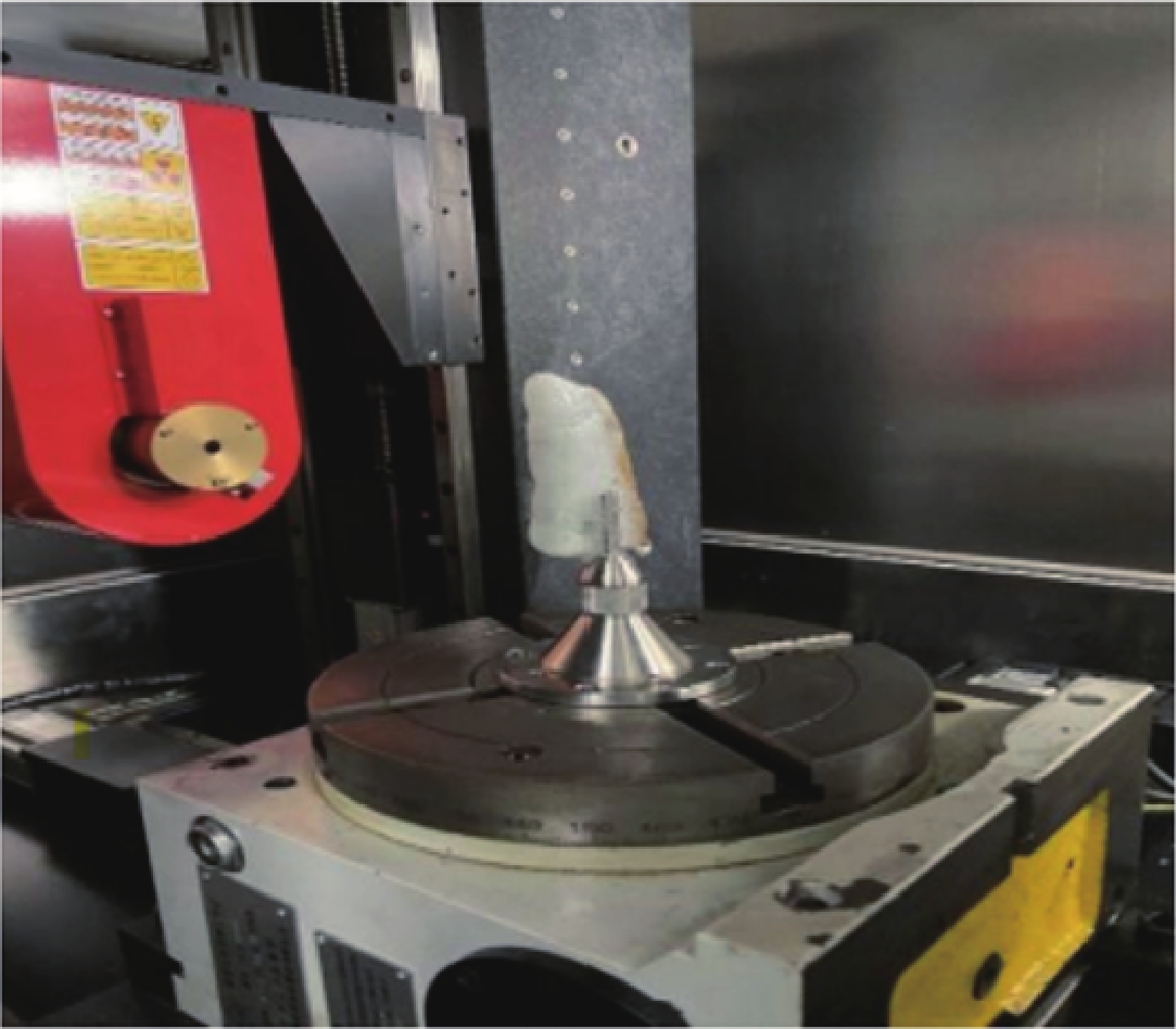
图 5 课题组使用CD-130BX/μCT微纳三维分析仪对岩矿样品进行扫描
Figure 5. The research group used the CD-130BX/μCT micro-nano 3D analyzer to scan rock and ore samples
表 1 工业CT总结
Table 1 Summary of industrial CT
CT种类 理论方法 传播速度km/s 操作难度 精度 经济成本 地震波CT 射线理论 5.5-7 中 高 高 电磁波CT 射线理论 约3×10⁵ 中 较高 中 电阻率CT 高密度电法 — 易 中 低  下载: 导出CSV
表 2 CT技术在研究不同材料孔隙结构中的应用
Table 2 Application of CT technology to study the pore structure of various materials
孔隙结构 孔隙半径R/μm 使用CT种类 结论 页岩孔隙结构 4~40×10-3 X-CT 岩心不同部位形成不同数量的孔隙空间 煤岩孔隙结构 0.1~100 X-CT 孔隙结构与煤岩的体积分形维数有关 黄土孔隙结构 2~6 Micro-CT 孔隙体的渗透率随孔隙度的增大而增大
下载: 导出CSV
-
[1] 高丽娜, 陈文革. CT技术的应用发展及前景[J]. CT理论与应用研究, 2009,18(1): 99−109. GAO L N, CHEN W G. The application and prospect of CT[J]. CT Theory and Applications, 2009, 18(1): 99−109. (in Chinese).
[2] 顾孝同. 国内工程CT技术的发展与应用[J]. 工程地球物理学报, 2006,3(4): 278−282. DOI: 10.3969/j.issn.1672-7940.2006.04.007. GU X T. Developments and applications of engineering CT technologies[J]. Chinese Journal of Engineering Geophysics, 2006, 3(4): 278−282. DOI: 10.3969/j.issn.1672-7940.2006.04.007. (in Chinese).
[3] CORMACK A M. Representation of a function by its line integrals, with some radiological applications[J]. Journal of Applied Physics, 1963, 34(9): 2722−2727. DOI: 10.1063/1.1729798.
[4] 庄天戈. 医用X-线成像历史的追溯、思考与期盼−为纪念我国第一台CT诞生30周年而作[J]. 中国医疗器械杂志, 2013,37(6): 391−394. doi: 10.3969/j.issn.1671-7104.2013.06.001 [5] 王革. X射线成像和深度学习的交叉融合[J]. CT理论与应用研究, 2022,31(1): 1−12. DOI: 10.15953/j.ctta.2021.053. WANG G. X-ray imaging meets deep learning[J]. CT Theory and Applications, 2022, 31(1): 1−12. DOI: 10.15953/j.ctta.2021.053. (in Chinese).
[6] 倪培君, 李旭东, 彭建中. 工业CT技术[J]. 无损检测, 1996,18(6): 173−176. NI P J, LI X D, PENG J Z. Industrial CT technique[J]. Nondestructive Testing, 1996, 18(6): 173−176. (in Chinese).
[7] 卢艳平, 王珏, 喻洪麟. 工业CT三维图像处理与分析系统[J]. 仪器仪表学报, 2009,30(2): 444−448. DOI: 10.19650/j.cnki.cjsi.2009.02.041. LU Y P, WANG J, YU H L. 3D image processing and analyzing system for industrial computed tomography[J]. Chinese Journal of Scientific Instrument, 2009, 30(2): 444−448. DOI: 10.19650/j.cnki.cjsi.2009.02.041. (in Chinese).
[8] 方黎勇, 李柏林, 李辉, 等. 工业CT在反求工程上的应用[J]. 强激光与粒子束, 2013,25(7): 1620−1624. DOI: 10.3788/HPLPB20132507.1620. FANG L Y, LI B L, LI H, et al. Application of industrial CT in reverse engineering technology[J]. High Power Laser and Particle Beams, 2013, 25(7): 1620−1624. DOI: 10.3788/HPLPB20132507.1620. (in Chinese).
[9] 张鹰, 郑红岩, 张志芳. 奋发图强, 为振兴中华而战−中国第一台γ射线 ICT实用样机诞生记[J]. 中国高等教育, 1995,(1): 39−41. [10] 段黎明, 刘元宝, 吴志芳, 等. 基于工业计算机断层成像技术的三维CAD模型重构方法[J]. 计算机集成制造系统, 2009,15(3): 479−486. DOI: 10.13196/j.cims.2009.03.65.duanlm.015. DUAN L M, LIU Y B, WU Z F, et al. Method of reconstructing 3-D CAD model based on industrial computed tomography[J]. Computer Integrated Manufacturing Systems, 2009, 15(3): 479−486. DOI: 10.13196/j.cims.2009.03.65.duanlm.015. (in Chinese).
[11] 孙晶晶, 杨民, 刘静华. 涡轮叶片CT图像边缘提取的最优算子研究[J]. 航空动力学报, 2010,25(1): 175−179. DOI: 10.13224/j.cnki.jasp.2010.01.038. SUN J J, YANG M, LIU J H. Research on optimal operator of blade edge detection from computed tomography images based on computational theory[J]. Journal of Aerospace Power, 2010, 25(1): 175−179. DOI: 10.13224/j.cnki.jasp.2010.01.038. (in Chinese).
[12] 胡俊杰, 徐洪苗, 王鹏, 等. 基于三维可视化的跨孔电磁波CT在岩溶勘察方面的应用[J]. 工程地球物理学报, 2022, 19(4): 443−449. DOI: 10.3969/j.issn.1672-7940.2022.04.004. HU J J, XU H M, WANG P, et al. Application of cross-hole electromagnetic wave tomography based on 3D visualization in Karst exploration[J]. Chinese Journal of Engineering Geophysics, 2022, 19(4): 443−449. DOI:10.3969/j.issn.1672-7940.2022.04.004. (in Chinese).
[13] 许继峰, 储著银, 李杰, 等. 难熔元素和代表性放射性同位素体系分析技术的进展、问题和应用展望[J]. 岩石学报, 2022,38(6): 1565−1576. DOI: 10.18654/1000-0569/2022.06.01. XU J F, CHU Z Y, LI J, et al. Progress, open questions and application prospects of analytical techniques for refractory elements and representative radioisotope systems[J]. Acta Petrologica Sinica, 2022, 38(6): 1565−1576. DOI: 10.18654/1000-0569/2022.06.01. (in Chinese).
[14] 田德祥, 刘新利, 王德志. 超声波在材料工程中的应用研究进展[J]. 材料研究与应用, 2022,16(6): 942−958. DOI: 10.20038/j.cnki.mra.2022.000608. TIAN D X, LIU X L, WANG D Z. Research progress of ultrasonic technology in materials engineering applications[J]. Materials Research and Application, 2022, 16(6): 942−958. DOI: 10.20038/j.cnki.mra.2022.000608. (in Chinese).
[15] 邹子龙. 地震CT技术及其在工程地质勘探中的应用[J]. 有色金属设计, 2020,47(3): 109−111. doi: 10.3969/j.issn.1004-2660.2020.03.032 ZOU Z L. Seismic CT technique and its application in engineering geological exploration[J]. Nonferrous Metals Design, 2020, 47(3): 109−111. (in Chinese). doi: 10.3969/j.issn.1004-2660.2020.03.032
[16] 童平. 地震层析成像方法及其应用研究[D]. 北京: 清华大学, 2012. [17] 陈敬. 基于波形反演的地震定位和层析成像研究[D]. 北京: 清华大学, 2021. DOI: 10.27266/d.cnki.gqhau.2021.000136. [18] TAPE C, LIU Q, MAGGI A, et al. Adjoint tomography of the southern California crust[J]. Science, 2009, 325(5943): 988−992. DOI: 10.1126/science.1175298.
[19] LIU Q, GU Y J. Seismic imaging: From classical to adjoint tomography[J]. Tectonophysics, 2012, 566: 31−66. DOI: 10.1016/j.tecto.2012.07.006.
[20] 张超, 刘伟, 褚金桥, 等. 电磁波 CT 技术在工程地质勘察中的应用[J]. Geomatics Science and Technology, 2020,8(2): 47−53. DOI: 10.12677/GST.2020.82006. ZHANG C, LIU W, CHU J Q, et al. Application of electromagnetic wave CT technology in engineering geological survey[J]. Geomatics Science and Technology, 2020, 8(2): 47−53. DOI: 10.12677/GST.2020.82006. (in Chinese).
[21] 陈杭, 邢亚东, 邓超云. 跨孔电磁波CT在煤矿采空区探测中的应用探究[J]. 矿山测量, 2022,50(1): 21−23, 37. DOI: 10.3969/j.issn.1001-358X.2022.01.006. CHEN H, XING Y D, DENG C Y. Application of borehole electromagnetic wave CT in coal mine goaf detection[J]. Mine Surveying, 2022, 50(1): 21−23, 37. DOI: 10.3969/j.issn.1001-358X.2022.01.006. (in Chinese).
[22] 郭光裕. 无线电波坑道透视技术在煤矿勘探隐伏地质构造中的应用[J]. 煤炭技术, 2018,37(8): 116−118. DOI: 10.13301/j.cnki.ct.2018.08.044. GUO G Y. Application of radio wave tunnel perspective technology in exploration of concealed geological structure in coal mine[J]. Coal Technology, 2018, 37(8): 116−118. DOI: 10.13301/j.cnki.ct.2018.08.044. (in Chinese).
[23] HE T, LI G, LUO F, et al. Research on mining-induced stress distribution of extrathick coal seams based on electromagnetic wave CT technology[J]. Advances in Civil Engineering, 2022, 2022. DOI: 10.1155/2022/6870207.
[24] HUANG S, LIN J, HUANG Q, et al. An emerging method using electromagnetic wave computed tomography for the detection of Karst caves[J]. Geotechnical and Geological Engineering, 2020, 38: 2713−2723. DOI: 10.1007/s10706-019-01180-w.
[25] 甘满光, 缪秀秀, 张力为, 等. CT扫描技术在二氧化碳地质利用与封存领域的应用研究综述[J]. 水利水电技术, 2019,50(8): 174−184. DOI: 10.13928/j.cnki.wrahe.2019.08.022. GAN M G, MIAO X X, ZHANG L W, et al. Review on applications of CT scanning technique in the field of CO2 geological utilization and storage[J]. Water Resources and Hydropower Engineering, 2019, 50(8): 174−184. DOI: 10.13928/j.cnki.wrahe.2019.08.022. (in Chinese).
[26] 张小海, 刘二军. 射线照相检验中X射线强度衰减的数值分析[J]. 无损检测, 2013,34(6): 1−4. DOI: cnki:sun:wsjc.0.2012-06-004. ZHANG X H, LIU E J. Numerical analysis on X-ray intensity attenuation in radiographic testing[J]. Nondestructive Testing, 2013, 34(6): 1−4. DOI: cnki:sun:wsjc.0.2012-06-004. (in Chinese).
[27] YAN C H, WHALEN R T, BEAUPRE G S, et al. Reconstruction algorithm for polychromatic CT imaging: Application to beam hardening correction[J]. IEEE Transactions on Medical Imaging, 2000, 19(1): 1−11. DOI: 10.1109/42.832955.
[28] 魏雨浓, 石战结, 余天祥. 不同电极阵列联合反演在古墓探测中的应用[J]. CT理论与应用研究, 2022,31(3): 280−292. DOI: 10.15953/j.ctta.2022.008. WEI Y N, SHI Z J, YU T X. Application of joint inversion of different electrode arrays in ancient mausoleum detection[J]. CT Theory and Applications, 2022, 31(3): 280−292. DOI: 10.15953/j.ctta.2022.008. (in Chinese).
[29] 方易小锁, 孟永东, 田斌, 等. 高密度电阻率法对不同电极排列的分辨率响应研究[J]. 地球物理学进展, 2019,34(6): 2424−2428. DOI: 10.6038/pg2019CC0313. FANGYI X S, MENG Y D, TIAN B, et al. Study on resolution response of different electrode arrangements by high density resistivity method[J]. Progress in Geophysic, 2019, 34(6): 2424−2428. DOI: 10.6038/pg2019CC0313. (in Chinese).
[30] BING Z, GREENHALGH S A. Cross-hole resistivity tomography using different electrode configurations[J]. Geophysical Prospecting, 2000, 48(5): 887−912. DOI: 10.1046/j.1365-2478.2000.00220.x.
[31] MOREIRA C A, GUIRELI NETTO L, CAMARERO P L, et al. Application of electrical resistivity tomography (ERT) in uranium mining earth dam[J]. Journal of Geophysics and Engineering, 2022, 19(6): 1265−1279. DOI: 10.1093/jge/gxac082.
[32] YI M, KIM J, SON J. Three-dimensional anisotropic inversion of resistivity tomography data in an abandoned mine area[J]. Exploration Geophysics, 2011, 42(1): 7−17. DOI: 10.1071/EG11005.
[33] NIELSON T, BRADFORD J, PIERCE J, et al. Soil structure and soil moisture dynamics inferred from time-lapse electrical resistivity tomography[J]. Catena, 2021, 207: 105553. DOI: 10.1016/j.catena.2021.105553.
[34] de JONG S M, HEIJENK R A, NIJLAND W, et al. Monitoring soil moisture dynamics using electrical resistivity tomography under homogeneous field conditions[J]. Sensors, 2020, 20(18): 5313. DOI: 10.3390/s20185313.
[35] GAO W, SHI L, ZHAI P. Water detection within the working face of an underground coal mine using 3D electric resistivity tomography (ERT)[J]. Journal of Environmental and Engineering Geophysics, 2019, 24(3): 497−505. DOI: 10.2113/JEEG24.3.497.
[36] CHENG Q, CHEN X, TAO M, et al. Characterization of Karst structures using quasi-3D electrical resistivity tomography[J]. Environmental Earth Sciences, 2019, 78: 1−12. DOI: 10.1007/s12665-019-8284-2.
[37] WANG Y, ANDERSON N, TORGASHOV E. Condition assessment of building foundation in Karst terrain using both electrical resistivity tomography and multi-channel analysis surface wave techniques[J]. Geotechnical and Geological Engineering, 2020, 38: 1839−1855. DOI: 10.1007/s10706-019-01133-3.
[38] 朱飞飞. 地井联合物探技术在岩溶注浆检测中的应用[J]. 工程地球物理学报, 2022,19(4): 450−458. DOI: 10.3969/j.issn.1672-7940.2022.04.005. ZHU F F. Application of geophysical prospecting technology combined with ground and well in Karst grouting detection[J]. Chinese Journal of Engineering Geophysics, 2022, 19(4): 450−458. DOI: 10.3969/j.issn.1672-7940.2022.04.005. (in Chinese).
[39] 车传强, 陈波, 谢明佐, 等. 综合物探方法在高压架空线路下方采空区探测中的应用[J]. CT理论与应用研究, 2022,31(1): 23−31. DOI: 10.15953/j.1004-4140.2022.31.01.03. CHE C Q, CHEN B, XIE M Z, et al. Application of integrated geophysics method in goaf detection under high voltage overhead lines[J]. CT Theory and Applications, 2022, 31(1): 23−31. DOI: 10.15953/j.1004-4140.2022.31.01.03. (in Chinese).
[40] LU Y, CAO C, LIU Y, et al. Study on application of comprehensive geophysical prospecting method in urban geological survey-taking concealed bedrock detection as an example in dingcheng district, Changde city, Hunan province, China[J]. Applied Sciences-basel, 2023, 13(1): 417. DOI: 10.3390/app13010417.
[41] ZHANG L, XU L, XIAO Y, et al. Application of comprehensive geophysical prospecting method in water accumulation exploration of multilayer goaf in integrated mine[J]. Advances in Civil Engineering, 2021, 2021: 1434893. DOI: 10.1155/2021/1434893.
[42] 朱亚军, 王艳新. 高密度电法和瞬变电磁法在地下岩溶探测中的综合应用[J]. 工程地球物理学报, 2012,9(6): 738−742. DOI: 10.3969/j.issn.1672-7940.2012.06.017. ZHU Y J, WANG Y X. The application of high density resistivity and TEM method to underground Karst detection[J]. Chinese Journal of Engineering Geophysics, 2012, 9(6): 738−742. DOI: 10.3969/j.issn.1672-7940.2012.06.017. (in Chinese).
[43] 杨峰, 刘晓甲, 李鹏博. 综合评定法在西南地区铁路岩溶路基注浆质量检测的应用[J]. 工程地球物理学报, 2021,15(8): 744−753. DOI: 10.3969/j.issn.1672-7940.2021.05.027. YANG F, LIU X J, LI P B. Application of comprehensive evaluation method in grouting quality detection of railway Karst subgrade in Southwest China[J]. Chinese Journal of Engineering Geophysics, 2021, 15(8): 744−753. DOI: 10.3969/j.issn.1672-7940.2021.05.027. (in Chinese).
[44] 章飞亮, 田占峰. 综合物探技术在空铁岩溶探测中的应用[J]. 工程地球物理学报, 2021,18(5): 730−737. DOI: 10.3969/j.issn.1672-7940.2021.05.025. ZHANG F L, TIAN Z F. Application of comprehensive geophysical technology in air-rail Karst detection[J]. Chinese Journal of Engineering Geophysics, 2021, 18(5): 730−737. DOI: 10.3969/j.issn.1672-7940.2021.05.025. (in Chinese).
[45] 唐塑, 武银婷, 邢浩, 等. 高密度电法与瞬变电磁法在戈壁区找水的联合应用[J]. CT理论与应用研究, 2023,32(1): 27−34. DOI: 10.15953/j.ctta.2022.081. TANG S, WU Y T, XING H, et al. Combined application of high-density electrical method and transient electromagnetic method in Gobi desert area[J]. CT Theory and Applications, 2023, 32(1): 27−34. DOI: 10.15953/j.ctta.2022.081. (in Chinese).
[46] 余涛, 王小龙, 王俊超. 综合物探方法在城市地铁岩溶勘察中的应用[J]. CT理论与应用研究, 2022,31(5): 587−596. DOI: 10.15953/j.ctta.2021.073. YU T, WANG X L, WANG J C. Application of comprehensive geophysical prospecting method in Karst exploration of urban subway[J]. CT Theory and Applications, 2022, 31(5): 587−596. DOI: 10.15953/j.ctta.2021.073. (in Chinese).
[47] 胡富彭, 欧元超, 付茂如. 不同充填介质下的溶洞跨孔电阻率CT探查数值模拟[J]. 中国岩溶, 2019,38(5): 766−773. DOI: 10.11932/Karst20190513. HU F P, OU Y C, FU M R. Study on numerical simulation of Karst cross-hole resistivity CT exploration at cave with different filling media[J]. Carsologica Sinica, 2019, 38(5): 766−773. DOI: 10.11932/Karst20190513. (in Chinese).
[48] TSOURLOS P, OGILVY R, PAPAZACHOS C, et al. Measurement and inversion schemes for single borehole-to-surface electrical resistivity tomography surveys[J]. Journal of Geophysics and Engineering, 2011, 8(4): 487−497. DOI: 10.1088/1742-2132/8/4/001.
[49] 周权, 王莉蓉. 地球物理方法在金属矿深部找矿中的应用及展望[J]. 世界有色金属, 2021,(9): 49−50. DOI: 10.3969/j.issn.1002-5065.2021.09.025. ZHOU Q, WANG L R. Application and prospect of geophysical methods in deep prospecting of metal deposits[J]. World Nonferrous Metals, 2021, (9): 49−50. DOI: 10.3969/j.issn.1002-5065.2021.09.025. (in Chinese).
[50] MOJICA A, PÉREZ T, TORAL J, et al. Shallow electrical resistivity imaging of the limón fault, Chagres river watershed, Panama Canal[J]. Journal of Applied Geophysics, 2017, 138: 135−142. DOI: 10.1016/j.jappgeo.2017.01.010.
[51] JUNG H B, JANSIK D, UM W. Imaging wellbore cement degradation by carbon dioxide under geologic sequestration conditions using X-ray computed microtomography[J]. Environmental science & technology, 2013, 47(1): 283−289. DOI: 10.1021/es3012707.
[52] 高星. 地震层析成像研究的回顾与展望[J]. 地球物理学进展, 2000,15(4): 41−45. DOI: 10.3969/j.issn.1004-2903.2000.04.006. GAO X. Advance and review in seismic tomography research[J]. Progress in Geophysics, 2000, 15(4): 41−45. DOI: 10.3969/j.issn.1004-2903.2000.04.006. (in Chinese).
[53] ZHANG P, LEE Y I, ZHANG J. A review of high-resolution X-ray computed tomography applied to petroleum geology and a case study[J]. Micron, 2019, 124: 102702. DOI: 10.1016/j.micron.2019.102702.
[54] GALLMEISTER K, AZZAM R. Applications of X-ray computed tomography (CT) in engineering geology[J]. Advances in X-ray Tomography for Geomaterials, 2006: 135-142.
[55] 李国荣, 李永耀. 工程CT技术的发展与应用[J]. 延安职业技术学院学报, 2012,26(3): 90−91. doi: 10.3969/j.issn.1674-6198.2012.03.037 [56] 重庆真测科技股份有限公司. 纳米CT[EB/OL]. (2020-09-09)[2023-5-11]. http://www.zcict.com/. [57] 陈超, 魏彪, 梁婷, 等. 一种基于工业CT技术的岩芯样品孔隙度测量分析方法[J]. 物探与化探, 2013,37(3): 500−507. DOI: 10.11720/j.issn.1000-8918.2013.3.22. CHEN C, WEI B, LIANG T, et al. The application of industrial computation tomography (CT) to the analysis of core sample porosity[J]. Geophysical and Geochemical Exploration, 2013, 37(3): 500−507. DOI: 10.11720/j.issn.1000-8918.2013.3.22. (in Chinese).
[58] 张向东, 王浩, 敬鹏飞. 基于岩石“等效损伤”探究宏观断裂规律[J]. 中国地质灾害与防治学报, 2020,31(3): 117−125. DOI: 10.16031/j.cnki.issn.1003-8035.2020.03.16. ZHANG X D, WANG H, JING P F. Studying the macroscopic fracture rule based on rock "equivalent damage"[J]. The Chinese Journal of Geological Hazard and Control, 2020, 31(3): 117−125. DOI: 10.16031/j.cnki.issn.1003-8035.2020.03.16. (in Chinese).
[59] 宋力, 魏赛平, 谷麟, 等. 微孔洞缺陷岩石轴压下弹塑脆性模型损伤研究[J]. 有色金属(矿山部分), 2014,66(3): 59−63. DOI: 10.3969/j.issn.1671-4172.2014.03.016. SONG L, WEI S P, GU L, et al. Study on elastoplastic-brittle model damage under axial compression in rocks with micro-cavity defect[J]. Nonferrous Metals (Mine Section), 2014, 66(3): 59−63. DOI: 10.3969/j.issn.1671-4172.2014.03.016. (in Chinese).
[60] 古启雄, 黄震, 钟文, 等. 高温循环后花岗岩孔隙结构与物理力学特性演化规律研究[J]. 岩石力学与工程学报, 2022: 1-16. DOI: 10.13722/j.cnki.jrme.2022.1024. GU Q X, HUANG Z, ZHONG W, et al. Study on the variations of pore structure and physical and mechanical properties of granite after high temperature cycling[J]. Chinese Journal of Rock Mechanics and Engineering, 2022: 1-16. DOI:10.13722/j.cnki.jrme.2022.1024. (in Chinese).
[61] FAN L F, GAO J W, WU Z J, et al. An investigation of thermal effects on micro-properties of granite by X-ray CT technique[J]. Applied Thermal Engineering, 2018, 140: 505−519. DOI: 10.1016/j.applthermaleng.2018.05.074.
[62] 李晓雪, 郤保平, 何水鑫, 等. 热冲击下花岗岩的细观变化规律[J]. 矿业研究与开发, 2021,41(5): 67−73. DOI: 10.13827/j.cnki.kyyk.2021.05.013. LI X X, XI B P, HE S X, et al. Mcroscopic change laws of granite under thermal shock[J]. Mining Research and Development, 2021, 41(5): 67−73. DOI: 10.13827/j.cnki.kyyk.2021.05.013. (in Chinese).
[63] VIDANA PATHIRANAGEI S, GRATCHEV I, SOKOLOWSKI K A. Investigation of the microstructural characteristics of heated sandstone by micro-computed tomography technique[J]. Environmental Earth Sciences, 2022, 81(15): 401. DOI: 10.1007/s12665-022-10514-6.
[64] SAXENA N, HOWS A, HOFMANN R, et al. Rock properties from micro-CT images: Digital rock transforms for resolution, pore volume, and field of view[J]. Advances in Water Resources, 2019, 134: 103419. DOI: 10.1016/j.advwatres.2019.103419.
[65] 左顺吉, 冯鹏, 黄盼, 等. 基于双域自适应网络的岩矿样工业CT图像金属伪影校正算法研究[J]. CT理论与应用研究, 2022,31(6): 783−792. DOI: 10.15953/j.ctta.2022.041. ZUO S J, FENG P, HUANG P, et al. Metal artifact reduction algorithm for CT images of rock and mineral samples based on dual-domain adaptive network[J]. CT Theory and Applications, 2022, 31(6): 783−792. DOI: 10.15953/j.ctta.2022.041. (in Chinese).
[66] ANOVITZ L M, COLE D R. Characterization and analysis of porosity and pore structures[J]. Reviews in Mineralogy and geochemistry, 2015, 80: 61−164. DOI: 10.2138/rmg.2015.80.04.
[67] TIWARI P, DEO M, LIN C L, et al. Characterization of oil shale pore structure before and after pyrolysis by using X-ray micro CT[J]. Fuel, 2013, 107: 547−554. DOI: 10.1016/j.fuel.2013.01.006.
[68] WANG G, SHEN J, LIU S, et al. Three-dimensional modeling and analysis of macro-pore structure of coal using combined X-ray CT imaging and fractal theory[J]. International Journal of Rock Mechanics and Mining Sciences, 2019, 123: 104082. DOI: 10.1016/j.ijrmms.2019.104082.
[69] 王超, 吴丰, 陈义国, 等. 基于微米CT技术的黄土岩孔隙保存机制及渗透率各向异性[J]. 科学技术与工程, 2021,21(27): 11527−11535. DOI: 10.3969/j.issn.1671-1815.2021.27.010. WANG C, WU F, CHEN Y G, et al. Preservation mechanism of pores and permeability anisotropy of loessite based on micro-CT technology[J]. Science Technology and Engineering, 2021, 21(27): 11527−11535. DOI: 10.3969/j.issn.1671-1815.2021.27.010. (in Chinese).
[70] LI P, SHAO S. Can X-ray computed tomography (CT) be used to determine the pore-size distribution of intact loess?[J]. Environmental Earth Sciences, 2020, 79: 29. DOI: 10.1007/s12665-019-8777-z.
[71] 徐文世, 于兴河, 刘妮娜, 等. 天然气水合物开发前景和环境问题[J]. 天然气地球科学, 2005,16(5): 680−683. DOI: cnki:sun:tdkx.0.2005-05-029. XU W S, YU X H, LIU N N, et al. The development perapective and environmental problems of natural gas hydrates[J]. Natural Gas Geoscience, 2005, 16(5): 680−683. DOI: cnki:sun:tdkx.0.2005-05-029. (in Chinese).
[72] WATANABE S, SAITO K, OHMURA R. Crystal growth of clathrate hydrate in liquid water saturated with a simulated natural gas[J]. Crystal Growth & Design, 2011, 11(7): 3235−3242. DOI: 10.1021/cg2005024.
[73] 李彦龙, 刘昌岭, 刘乐乐. 含水合物沉积物损伤统计本构模型及其参数确定方法[J]. 石油学报, 2016,37(10): 1273−1279. DOI: 10.7623/syxb201610007. LI Y L, LIU C L, LIU L L. Damage statistic constitutive model of hydrate-bearing sediments and the determination method of parameters[J]. Acta Petrolei Sinica, 2016, 37(10): 1273−1279. DOI: 10.7623/syxb201610007. (in Chinese).
[74] LIU L, DAI S, NING F, et al. Fractal characteristics of unsaturated sands− implications to relative permeability in hydrate-bearing sediments[J]. Journal of Natural Gas Science and Engineering, 2019, 66: 11−17. DOI: 10.1016/j.jngse.2019.03.019.
[75] YANG L, AI L, XUE K, et al. Analyzing the effects of inhomogeneity on the permeability of porous media containing methane hydrates through pore network models combined with CT observation[J]. Energy, 2018, 163: 27−37. DOI: 10.1016/j.energy.2018.08.100.
[76] ZHANG L, GE K, WANG J, et al. Pore-scale investigation of permeability evolution during hydrate formation using a pore network model based on X-ray CT[J]. Marine and Petroleum Geology, 2020, 113: 104157. DOI: 10.1016/j.marpetgeo.2019.104157.
[77] 陈亮, 叶旺全, 李承峰, 等. 基于时间演化的天然气水合物CT图像阈值分割[J]. CT理论与应用研究, 2023,32(2): 171−178. DOI: 10.15953/j.ctta.2022.062. CHEN L, YE W Q, LI C F, et al. Natural gas hydrate CT image threshold segmentation based on time evolution[J]. CT Theory and Applications, 2023, 32(2): 171−178. DOI: 10.15953/j.ctta.2022.062. (in Chinese).
[78] 李小彬. 基于三维数字岩心的岩石孔隙结构表征及弹渗属性模拟研究[D]. 武汉: 中国地质大学(武汉), 2021. [79] ALHASHMI Z, BLUNT M J, BIJELJIC B. The impact of pore structure heterogeneity, transport, and reaction conditions on fluid–fluid reaction rate studied on images of pore space[J]. Transport in Porous Media, 2016, 115(2): 215−237. DOI: 10.1007/s11242-016-0758-z.
[80] LIN W, LI X, YANG Z, et al. Modeling of 3D rock porous media by combining X-ray CT and Markov chain Monte Carlo[J]. Journal of Energy Resources Technology, 2020, 142(1): 13001. DOI: 10.1115/1.4045461.
[81] 方黎勇, 陈鹏, 陈浩. 一种基于工业CT图像的岩心孔隙率计算方法[J]. 强激光与粒子束, 2014,26(3): 246−250. DOI: 10.3788/HPLPB201426.034008. FANG L Y, CHEN P, CHEN H. A calculation method of core porosity based on industrial CT images[J]. High Power Laser and Particle Beams, 2014, 26(3): 246−250. DOI: 10.3788/HPLPB201426.034008. (in Chinese).
[82] 姚军, 赵秀才, 衣艳静, 等. 数字岩心技术现状及展望[J]. 油气地质与采收率, 2005,(6): 52−54. DOI: 10.3969/j.issn.1009-9603.2005.06.017. YAO J, ZHAO X C, YI Y J. et al. The current situation and prospect on digital core technology[J]. Petroleum Geology and Recovery Efficiency, 2005, (6): 52−54. DOI: 10.3969/j.issn.1009-9603.2005.06.017. (in Chinese).
[83] LIN W, LI X, YANG Z, et al. Construction of dual pore 3-D digital cores with a hybrid method combined with physical experiment method and numerical reconstruction method[J]. Transport in Porous Media, 2017, 120(1): 227−238. DOI: 10.1007/s11242-017-0917-x.
[84] 赵建鹏, 陈惠, 李宁, 等. 三维数字岩心技术岩石物理应用研究进展[J]. 地球物理学进展, 2020,35(3): 1099−1108. DOI: 10.6038/pg2020DD0486. ZHAO J P, CHEN H, LI N, et al. Research advance of petrophysical application based on digital core technology[J]. Progress in Geophysics, 2020, 35(3): 1099−1108. DOI: 10.6038/pg2020DD0486. (in Chinese).
[85] 邓世冠, 吕伟峰, 刘庆杰, 等. 利用CT技术研究砾岩驱油机理[J]. 石油勘探与开发, 2014,41(3): 330−335. DOI: 10.11698/PED.2014.03.08. DENG S G, LV W F, LIU Q J, et al. Research on displacement mechanism in conglomerate using CT scanning method[J]. Petroleum Exploration and Development, 2014, 41(3): 330−335. DOI: 10.11698/PED.2014.03.08. (in Chinese).
[86] 屈乐, 孙卫, 杜环虹, 等. 基于CT扫描的三维数字岩心孔隙结构表征方法及应用−以莫北油田116井区三工河组为例[J]. 现代地质, 2014,28(1): 190−196. DOI: 10.3969/j.issn.1000-8527.2014.01.020. QU L, SUN W, DU H H, et al. Characterization technique of pore structure by 3D digital core based on CT scanning and its application: An example from Sangonghe formation of 116 well field in Mobei oilfield[J]. Geoscience, 2014, 28(1): 190−196. DOI: 10.3969/j.issn.1000-8527.2014.01.020. (in Chinese).
[87] 盛军, 杨晓菁, 李纲, 等. 基于多尺度X-CT成像的数字岩心技术在碳酸盐岩储层微观孔隙结构研究中的应用[J]. 现代地质, 2019,33(3): 653−661. DOI: 10.19657/j.geoscience.1000-8527.2019.03.17. SHENG J, YANG X J, LI G, et al. Aplication of multiscale X-CT imaging digital core technique on observing micro-pore structure of carbonate reservoirs[J]. Geoscience, 2019, 33(3): 653−661. DOI: 10.19657/j.geoscience.1000-8527.2019.03.17. (in Chinese).
[88] 郝艳军, 杨顶辉. 二氧化碳地质封存问题和地震监测研究进展[J]. 地球物理学进展, 2012,27(6): 2369−2383. DOI: 10.6038/j.issn.1004-2903.2012.06.012. HAO Y J, YANG D H. Research progress of carbon dioxide capture and geologicalsequestration problem and seismic monitoring research[J]. Progress in Geophysics, 2012, 27(6): 2369−2383. DOI: 10.6038/j.issn.1004-2903.2012.06.012. (in Chinese).
[89] ANDREW M, BIJELJIC B, BLUNT M J. Pore-scale contact angle measurements at reservoir conditions using X-ray microtomography[J]. Advances in Water Resources, 2014, 68(2014): 24−31. DOI: 10.1016/j.advwatres.2014.02.014.
[90] SAHNI A, BURGER J, BLUNT M. Measurement of three phase relative permeability during gravity drainage using CT[C]//SPE/DOE Improved Oil Recovery Symposium, 1998. DOI: 10.2118/39655-MS.
[91] ZHANG Y, NISHIZAWA O, KIYAMA T, et al. Flow behaviour of supercritical CO2 and brine in Berea sandstone during drainage and imbibition revealed by medical X-ray CT images[J]. Geophysical Journal International, 2014, 197(3): 1789−1807. DOI: 10.1093/gji/ggu089.
[92] 施有志, 赵花丽, 黄钰琳, 等. 厦门地区孤石分布规律及对地铁工程的影响[J]. 地质与勘探, 2019,55(3): 862−869. DOI: 10.12134/j.dzykt.2019.03.018. SHI Y Z, ZHAO H L, HUANG Y L, et al. The distribution rule of the solitary stones of granite in Xiamen and its influence on metro engineering[J]. Geology and Exploration, 2019, 55(3): 862−869. DOI: 10.12134/j.dzykt.2019.03.018. (in Chinese).
[93] 李术才, 刘征宇, 刘斌, 等. 基于跨孔电阻率CT的地铁盾构区间孤石探测方法及物理模型试验研究[J]. 岩土工程学报, 2015,37(3): 446−457. DOI: 10.11779/CJGE201503008. LI S C, LIU Z Y, LIU B, et al. Boulder detection method for metro shield zones based on cross-hole resistivity tomography and its physical model tests[J]. Chinese Journal of Geotechnical Engineering, 2015, 37(3): 446−457. DOI: 10.11779/CJGE201503008. (in Chinese).
[94] 刘畅, 李振春, 曲英铭, 等. 地震层析成像方法综述[J]. 物探与化探, 2020,44(2): 227−234. DOI: 10.11720/wtyht.2020.1243. LIU C, LI Z C, QU Y M, et al. A review of seismic tomography methods[J]. Geophysical and Geochemical Exploration, 2020, 44(2): 227−234. DOI: 10.11720/wtyht.2020.1243. (in Chinese).
[95] 秦晶晶, 袁洪克, 何银娟, 等. 层析成像技术在城市活断层探测中的应用[J]. 地球物理学进展, 2018,33(5): 2153−2158. DOI: 10.6038/pg2018BB0383. QIN J J, YUAN H K, HE Y J, et al. Application of tomography inversion method in detecting active fault[J]. Progress in Geophysics, 2018, 33(5): 2153−2158. DOI: 10.6038/pg2018BB0383. (in Chinese).
[96] GHORBANI Y, BECKER M, PETERSEN J, et al. Use of X-ray computed tomography to investigate crack distribution and mineral dissemination in sphalerite ore particles[J]. Minerals Engineering, 2011, 24(12): 1249−1257. DOI: 10.1016/j.mineng.2011.04.008.
[97] LIU Z, YANG Y, YAO J, et al. Pore-scale remaining oil distribution under different pore volume water injection based on CT technology[J]. Advances in Geo-Energy Research, 2017, 1(3): 171−181. DOI: 10.26804/ager.2017.03.04.
[98] WILDENSCHILD D, SHEPPARD A P. X-ray imaging and analysis techniques for quantifying pore-scale structure and processes in subsurface porous medium systems[J]. Advances in Water Resources, 2013, 51: 217−246. DOI: 10.1016/j.advwatres.2012.07.018.
-
期刊类型引用(3)
1. 杨文杰,梁烨,杨柯. 双流法肺动脉技术在肺动脉扫描中的应用评价. 影像诊断与介入放射学. 2024(04): 278-282 .  百度学术
百度学术
2. 胡嘉诚,刘云福,王新艳,王倩,马梓轩,张永县,李伟,刘荣,牛延涛. 对比剂分次团注联合能量成像获得CTU和CTA联合成像的应用研究. CT理论与应用研究. 2024(06): 692-700 . 本站查看
3. 肖爱兰. 基于肺动脉CTA对肺栓塞检出流程优化的探讨. 现代医用影像学. 2024(09): 1644-1646+1656 . 百度学术
其他类型引用(0)


计量
- 文章访问数: 516
- HTML全文浏览量: 72
- PDF下载量: 87
- 被引次数: 3


