
Research on Image Analysis Method of Spectral CT Based on Principal Component Analysis
-
摘要: 基于光子计数探测器的能谱CT,可以同时采集多个能谱通道的投影数据,并获得相应能量范围内物质的吸收特征,可以有效应用于物质识别与材料分解。主成分分析是一种很好的多元数据分析技术,可以用于处理多能谱CT数据。本文分别在投影域和图像域对能谱CT数据进行主成分分析,并对分析结果做出系统比较。为了减少噪声的影响,提高能谱CT图像的彩色表征性能,提出双域滤波与像素值平方相结合的方法,用于含噪声的主成分图像去噪,然后将所选取的主成分图像映射到RGB颜色通道。实验结果表明,无论是在投影域还是图像域进行主成分分析,都可以获取清晰的CT图像,识别出物质的不同成分。相较于在图像域的主成分分析方法,在投影域进行主成分分析能够保留物质的更多细节,获取更清晰的彩色CT图像。Abstract: Spectral computed tomography (CT) based on photon counting detector can simultaneously collect projection data of multiple spectral channels and obtain absorption characteristics of material within corresponding energy ranges, so it can be effectively applied to material identification and material decomposition. Principal component analysis is an excellent multivariate analysis technique, which can be applied to process multi-energy spectral CT data. In this paper, principal component analysis was performed on spectral CT data in projection domain and image domain respectively, and the analysis results were compared systematically. Meanwhile, in order to reduce the influence of noise and improve the color characterization performance of spectral CT images, the method of combining double domain filtering with pixel value square was proposed to denoise the noisy principal component images, and then the selected principal component images were mapped to RGB color channels. The experimental results demonstrate that the principal component analysis can obtain clear CT images and identify the different components of the substance, whether in the projection domain or the image domain. However, compared with the principal component analysis method in the image domain, principal component analysis in the projection domain can retain more details of the substance and acquire clearer color CT images.
-
-
![]()
图 4 小鼠胸腔投影域的主成分分析图像
Figure 4. PCA images of projection domain of mouse thoracic cavity
![]()
图 5 小鼠胸腔投影域主成分分析重建图像
Figure 5. Reconstructed images of projection domain of mouse thoracic cavity by PCA
![]()
图 6 小鼠胸腔图像域的主成分分析图像
Figure 6. PCA images of the image domain of the mouse thoracic cavity
![]()
图 7 小鼠胸腔的像素值平方去噪的主成分分析图像
Figure 7. PCA image of pixel value square denoising of mouse thoracic cavity
![]()
图 8 小鼠胸腔的双域滤波与像素值平方相结合去噪的主成分分析图像
Figure 8. PCA image denoising combining dual-domain filtering with pixel value square of mouse thoracic cavity
![]()
图 13 临床前小鼠投影域的主成分分析重建图像
Figure 13. Reconstructed images of projection domain preclinical mice by PCA
![]()
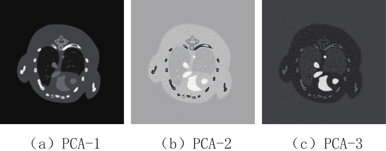
图 14 临床前小鼠图像域的主成分分析图像
Figure 14. PCA images of the image domain of the preclinical mice
![]()
图 15 临床前小鼠投影域第2主成分重建图像的去噪图像
Figure 15. Denoised images of the PCA-2 recon-structed images in the projection domain of preclinical mice
![]()
图 16 临床前小鼠图像域第2主成分的去噪图像
Figure 16. Denoised images of PCA-2 in the image domain of preclinical mice
表 1 小鼠胸腔投影域与图像域进行主成分分析的各个主成分的贡献率
Table 1 The contribution rate of each principal component of the projection domain and image domain of the mouse thoracic cavity for PCA
区域 各个主成分的贡献率/% PCA-1 PCA-2 PCA-3 PCA-4 PCA-5 PCA-6 PCA-7 PCA-8 投影域 99.758 0.146 0.071 0.007 0.005 0.005 0.004 0.004 图像域 98.094 1.234 0.591 0.026 0.020 0.014 0.012 0.009  下载: 导出CSV
表 2 两种去噪算法在小鼠胸腔投影域与图像域主成分图像上的峰值信噪比
Table 2 Peak signal to noise ratio of the two denoising algorithms for principal component images of mouse thoracic cavity in the projection domain and image domain
方法 投影域 图像域 PCA-2 PCA-3 PCA-2 PCA-3 像素值平方 64.209 49.139 51.590 61.749 双域滤波与像素值平方相结合 64.257 49.139 51.589 61.865
下载: 导出CSV
表 3 临床前小鼠在投影域与图像域进行主成分分析的各个主成分的贡献率
Table 3 The contribution rate of each principal component of the projection domain and image domain of the preclinical mice for PCA
区域 PCA-1 PCA-2 PCA-3 PCA-4 PCA-5 PCA-6 PCA-7 投影域PCA贡献率/% 99.727 0.060 0.024 0.020 0.020 0.019 0.019 图像域PCA贡献率/% 99.544 0.222 0.032 0.022 0.021 0.021 0.021 区域 PCA-8 PCA-9 PCA-10 PCA-11 PCA-12 PCA-13 投影域PCA贡献率/% 0.019 0.019 0.019 0.018 0.018 0.018 图像域PCA贡献率/% 0.020 0.020 0.020 0.020 0.019 0.018
下载: 导出CSV
-
[1] WU X C, HE P, ZHANG Y, et al. The small animal material discrimination study based on equivalent monochromatic energy projection decomposition method with dual-energy CT system[J]. Journal of X-ray Science and Technology, 2018, 26(6): 919−929. doi: 10.3233/XST-180418
[2] AARON S, SAVVAS N. Spectral computed tomography: Fundamental principles and recent developments[J]. Korean Journal of Radiology, 2021, 22(1): 86−96. doi: 10.3348/kjr.2020.0144
[3] FRÉDÉRIC J, CLARISSE F, MICHEL G, et al. An alternating projection-image domains algorithm for spectral CT[C]//Proceedings. IEEE International Symposium on Biomedical Imaging, United states, 2020: 187-190.
[4] GENG M F, TIAN Z F, JIANG Z, et al. PMS-GAN: Parallel multi-stream generative adversarial network for multi-material decomposition in spectral computed tomography[J]. IEEE Transactions on Medical Imaging, 2021, 40(2): 571−584. doi: 10.1109/TMI.2020.3031617
[5] LIU J L, DING H J, MOLLOI S, et al. TICMR: Total image constrained material reconstruction via nonlocal total variation regularization for spectral CT[J]. IEEE Transactions on Medical Imaging, 2016, 35(12): 2578−2586. doi: 10.1109/TMI.2016.2587661
[6] FENG J, YU H J, WANG S Y, et al. Image-domain based material decomposition by multi-constraint optimization for spectral CT[J]. IEEE Access, 2020, 8: 155450−155458. doi: 10.1109/ACCESS.2020.3016675
[7] WU W W, YU H J, CHEN P J, et al. Dictionary learning based image-domain material decomposition for spectral CT[J]. Physics in Medicine and Biology, 2020, 65(24): 245006. doi: 10.1088/1361-6560/aba7ce
[8] ABASCAL J F P J, DUCROS N, PEYRIN F. Nonlinear material decomposition using a regularized iterative scheme based on the bregman distance[J]. Inverse Problems, 2018, 34(12): 124003. doi: 10.1088/1361-6420/aae1e7
[9] 乔志伟. 总变差约束的数据分离最小图像重建模型及其Chambolle-Pock求解算法[J]. 物理学报, 2018,67(19): 383−396. doi: 10.7498/aps.67.20180839 QIAO Z W. The total variation constrained data divergence minimization model for image reconstruction and its Chambolle-Pock solving algorithm[J]. Acta Physica Sinica, 2018, 67(19): 383−396. (in Chinese). doi: 10.7498/aps.67.20180839
[10] KONG H H, LIU R, YU H Y. Ordered-subset split-bregman algorithm for interior tomography[J]. Journal of X-ray Science and Technology, 2016, 24(2): 221−240. doi: 10.3233/XST-160547
[11] BHAYANA R, PARAKH A, KAMBADAKONE A, et al. Material decomposition with dual- and multi-energy computed tomography[J]. MRS Communications, 2020, 10(4): 558−565. doi: 10.1557/mrc.2020.86
[12] ANTHONY B, JOCHEN B, NANETTE S, et al. Processing of spectral X-ray data with principal components analysis[J]. Nuclear Instruments and Methods in Physics Research, Section A: Accelerators, Spectrometers, Detectors and Associated Equipment, 2011, 633(S1): S140−S142.
[13] ANDERSON N G, BUTLER A P, SCOTT N J A, et al. Spectroscopic (multi-energy) CT distinguishes iodine and barium contrast material in MICE[J]. European Radiology, 2010, 20(9): 2126−2134. doi: 10.1007/s00330-010-1768-9
[14] HE P, YU H Y, THAYER P, et al. Preliminary experimental results from a MARS micro-CT system[J]. Journal of X-ray Science and Technology, 2012, 20(2): 199−211. doi: 10.3233/XST-2012-0329
[15] KONG H H, LIU R, PAN J X, et al. Evaluation of an analytic reconstruction method as a platform for spectral cone-beam CT[J]. IEEE Access, 2018, 6: 21314−21323. doi: 10.1109/ACCESS.2018.2820500
[16] XIE H Q, REN Y, LONG W T, et al. Principal component analysis in projection and image domains: Another form of spectral imaging in photon-counting CT[J]. IEEE Transactions on Biomedical Engineering, 2021, 68(3): 1074−1083. doi: 10.1109/TBME.2020.3013491
[17] 周志华. 机器学习[M]. 北京: 清华大学出版社, 2016. ZHOU Z H. Machine Learning[M]. Beijing: Tsinghua University Press, 2016.
[18] KNAUS C, ZWICKER M. Dual-domain image denoising[C]//2013 IEEE International Conference on Image Processing, ICIP 2013: Proceedings, Australia, 2013: 440-444.
[19] 孔慧华, 连祥媛, 陈平, 等. 基于能谱CT的材料组分彩色表征研究[J]. 光谱学与光谱分析, 2021,41(11): 3612−3617. KONG H H, LIAN X Y, CHEN P, et al. Research on color characterization of material components based on spectral CT[J]. Spectroscopy and Spectral Analysis, 2021, 41(11): 3612−3617. (in Chinese).
-
期刊类型引用(6)
1. 王昱,张慧敏,邓雪蓉,刘伟伟,陈璐,赵宁,张晓慧,宋志博,耿研,季兰岚,王玉,张卓莉. 尿枸橼酸定量检测在原发性痛风患者肾结石诊断中的应用价值. 北京大学学报(医学版). 2022(06): 1134-1140 .  百度学术
百度学术
2. 赵玲玲. 多层螺旋CT低剂量平扫在诊断肾及输尿管结石中的应用价值分析. 中国CT和MRI杂志. 2019(05): 116-118 . 百度学术
3. 周镇源,张俊文,刘志锋,蔡金辉,阮耀钦,郭栋华,刘庆余,徐金戈. 常规定量CT鉴别尿路结石成分的研究. CT理论与应用研究. 2019(03): 331-338 . 本站查看
4. 吕文选,王丽琴,胡云宇,王峰岩,张艾红,巴建. 非增强CT值在预测体外冲击波碎石术治疗肾结石的应用价值研究. 中国CT和MRI杂志. 2018(06): 77-80 . 百度学术
5. 李丽超,宫凤玲,于鹏,刘俊娥,周立娟,黄孝华. 常规CT鉴别诊断尿酸与非尿酸结石的价值. 广西医学. 2018(03): 284-286 . 百度学术
6. 陈艾,商亚军,陈英. 泌尿系结石的CT值与结石成分及易碎性之间的关系. 深圳中西医结合杂志. 2018(17): 11-13 . 百度学术
其他类型引用(5)


计量
- 文章访问数: 666
- HTML全文浏览量: 219
- PDF下载量: 245
- 被引次数: 11


