The CT and MRI Findings and Differential Diagnosis of Sebaceous Carcinoma of Eyelid and Basal Cell Carcinoma
-
摘要: 目的:探讨睑板腺癌(SC)与基底细胞癌(BCC)的CT及MRI表现,提高对二者的鉴别诊断能力。方法:回顾性分析经手术证实的14例SC和7例BCC患者的临床资料和影像表现。结果:在14例SC患者中6例行CT平扫检查,7例行MRI平扫检查,1例行CT与MRI检查;在7例BCC患者中1例行CT平扫检查,5例行MRI平扫检查,1例行CT与MRI检查。SC病例中92.9% 为女性(13/14),50% 发生于上睑(7/14),病灶形态多数呈环条状和结节状(12/14),半数病灶边界不清晰(7/14),病灶内常出现“弧形征”(5/14)和气体征(4/7),1例侵犯邻近眼眶组织;BCC病例男女患者比例相近,病灶全部位于下睑(7/7),近半数病灶形态呈结节状(3/7),边界多数较清晰(6/7),1例病灶内见钙化灶,未见病灶侵犯邻近组织。结论:SC与BCC在流行病学、发病部位、影像学特征等方面均有一定的差异,掌握二者鉴别要点可提高术前定性诊断准确性。Abstract: Objective: To improve the ability of differential diagnosis of sebaceous carcinoma of eyelid and basal cell carcinoma by investigating their CT and MRI findings. Methods: The clinical data and imaging findings of 14 patients with sebaceous carcinoma of eyelid and 7 patients with basal cell carcinoma confirmed by operation were retrospectively analyzed and compared. Results: Among the 14 patients with SC, 6 cases underwent CT examination, 7 cases underwent the MRI examination, and 1 case underwent both CT and MRI. Among the 7 patients with BCC, 1 case underwent CT examination, and 5 cases underwent the MRI plain scan, and 1 case underwent both CT and MRI. 92.9% of SC cases were female (13/14). 50% of SC were located in the upper eyelid, and most of them were in the shape of ring strip and nodule (12/14). Half of the lesions had unclear boundaries (7/14). The arc sign (5/14) and gas sign (4/7) often appeared in the lesions. One case invaded the adjacent orbital tissue. The proportion of male and female patients in BCC cases was similar. The lesions of BCC were all located in the lower eyelid (7/7). Nearly half of the shape of the lesions was nodular (3/7). Most of them had clear boundaries (6/7). Calcification was found in one case, and no invasion of adjacent tissue was found. Conclusion: There are some differences in epidemiology, location and imaging features between sebaceous carcinoma of eyelid and basal cell carcinoma. Mastering the key points of differentiation between sebaceous carcinoma of eyelid and basal cell carcinoma can improve the accuracy of preoperative qualitative diagnosis.
-
基底细胞癌(basal cell carcinoma,BCC)与睑板腺癌(sebaceous carcinoma of eyelid,SC)是中老年人最常见的眼睑恶性肿瘤,发病率分别位于第1和第2位,但是因两者恶性程度和侵袭性的不同,患者的治疗方法和预后有很大不同[1-2]。术前对BCC和SC进行定性评估,对手术方法的选择、提高治愈率和降低转移率尤为重要。
本文回顾性分析我院2015年1月至2020年9月经手术病理证实的14例SC及7例BCC的影像学资料,对两种疾病的一般资料与CT、MRI表现做比较和总结,以提高对眼睑部疾病的认识和诊断准确率。
1. 资料和方法
1.1 一般资料
本组SC患者14例,男性1例,女性13例,年龄54~86岁,平均年龄(73.07±10.87)岁。BCC患者7例,男性3例,女性4例,年龄57~84岁,平均年龄(70.71±9.46)岁。临床症状均为发现眼睑部肿物或有异物感,2例既往有眼眶手术史。所有病例最终诊断均由手术病理证实。
1.2 检查方法
14例SC患者术前6例行CT平扫检查,7例行MRI平扫检查,1例行CT与MRI检查。7例BCC患者1例行CT平扫检查,5例行MRI平扫检查,1例行CT与MRI检查。
CT检查采用GE Light speed 16排CT扫描机。扫描参数:120 kV,200 mA,常规扫描层厚3 mm,薄层重建层厚1 mm,螺距1.0;MRI检查采用Philips 3.0 T扫描仪及GE 1.5 T MRI扫描仪,使用眼眶专用线圈,扫描序列包括T1 WI、T2 WI、T2抑脂,DWI,扫描方位包括横断位、冠状位、矢状位,扫描层厚4 mm。
1.3 图像分析
所有患者CT和MRI图像由两位具有5年以上头颈部影像诊断工作经验的放射诊断医师共同阅片,如有意见分歧,两位医师共同协商达成一致意见。分析内容包括病变部位、形态、大小、密度及信号特征、病灶与邻近结构的关系、有无钙化、气体、囊变等。
2. 结果
2.1 睑板腺癌
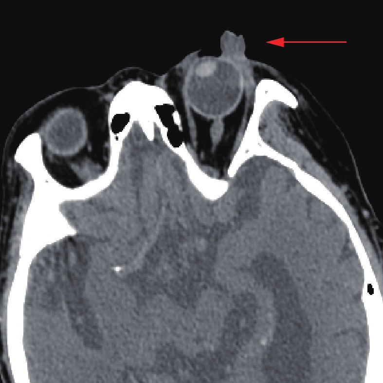
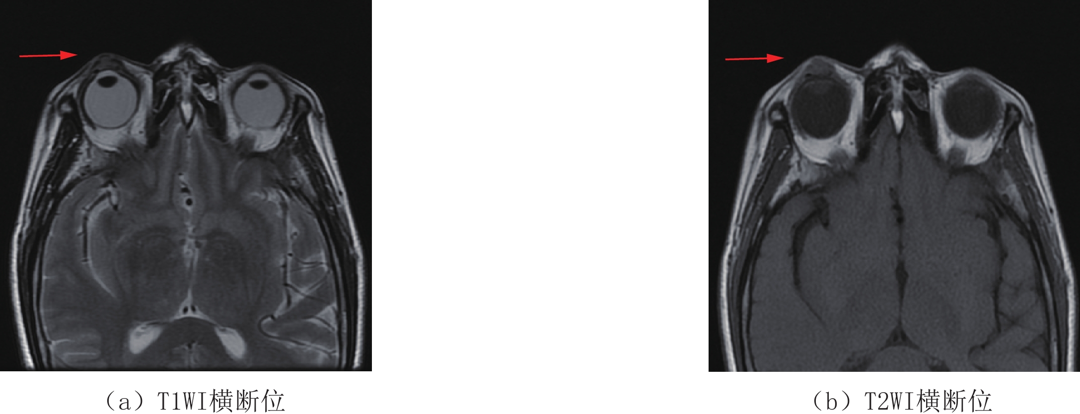
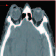
14例SC患者影像学表现。①发病部位:右眼睑6例,其中4例位于上睑,1例位于下睑,1例病变侵犯眶内;左眼睑8例,3例位于上睑,5例位于下睑。②形态及大小:14例肿瘤长径范围1.1~3.8 cm,平均长径(1.84±0.81)cm;5例肿瘤呈环条状及斑块状附着于眼环前方,可见“弧形征”(图1),8例肿瘤表现为眼睑部软组织结节,其中1例呈“菜花状”向外凸起(图2);1例肿瘤呈团块状向眼眶内生长;7例病灶边界不清晰,7例边界清晰。③病灶密度及信号特征:7例行CT检查的患者表现为等密度软组织影,病灶较大时密度不均匀,未见囊变坏死区;4例内部见气体密度影,均未见钙化影;8例行MRI检查的患者表现为T1 WI等或低信号,T2 WI呈稍高信号,脂肪抑制序列为高信号,DWI呈高或稍高信号。④病灶与邻近结构的关系:14例中13例局限于眶隔前区,11例表皮受累,2例仅位于皮下未侵及眼睑皮肤(图3),1例肿瘤范围较大,侵犯球内及球后,累及眼外肌、泪腺及视神经。
2.2 基底细胞癌
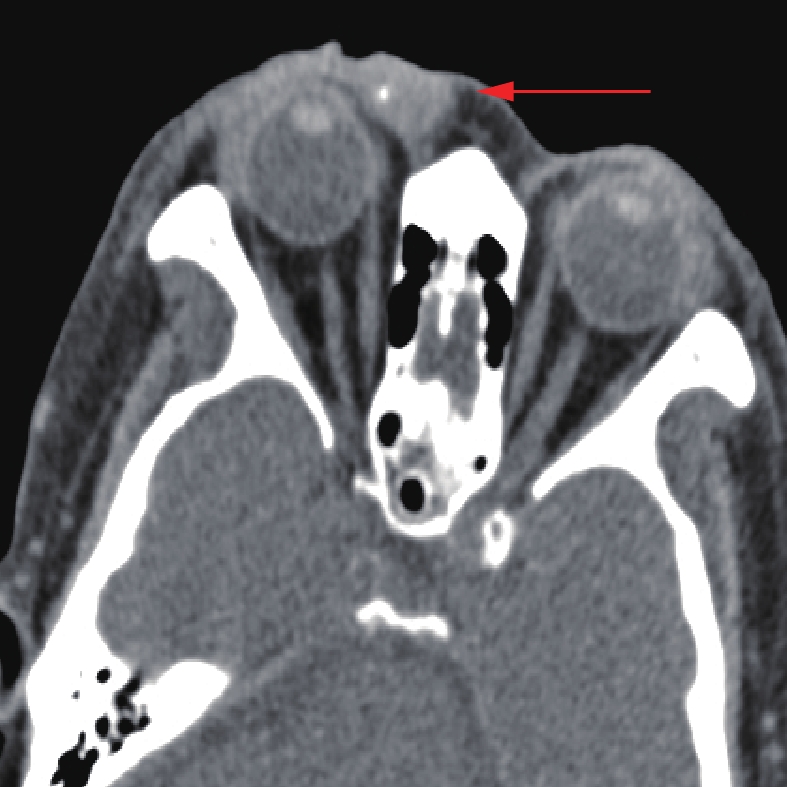
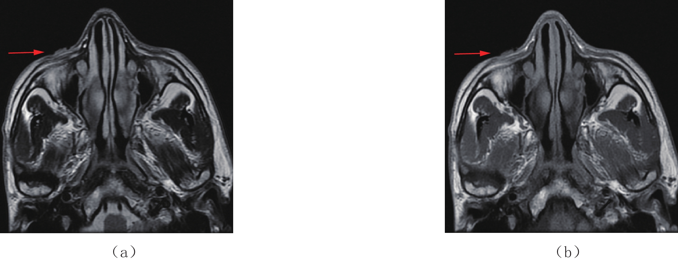
7例BCC患者影像学表现。①发病部位:右眼睑4例,左眼睑3例,均位于下睑。②形态及大小:1例呈环条状肿块,3例呈结节状,1例呈扁丘状,边界均较清晰;肿瘤长径范围0.6~4.4 cm,平均长径(1.60±1.58) cm;1例呈边界不清晰的团片状混杂信号影;1例仅见皮肤表面略不光整,未见明显肿块;仅1例见“弧形征”。③病灶密度及信号特征:2例行CT检查的患者表现为密度均匀的软组织影,1例病灶内见点状钙化(图4);6例行MRI检查的患者表现为T1 WI等或低信号,T2 WI呈稍高或高信号,DWI呈稍高信号。④病灶与邻近结构的关系:7例肿瘤均局限于眶隔前区,2例仅局限在皮肤,未侵犯皮下脂肪组织(图5)。
2.3 SC与BCC的鉴别诊断
SC与BCC的一般资料对比(表1)显示,女性患者比例SC(92.9%)高于BCC(57.1%)。
表 1 SC与BCC一般资料对比Table 1. The comparison of general information of SC and BCC肿瘤类型 例数 平均年龄/岁 性别 男 女 SC 14 73.07±10.87 1 13 BCC 7 70.71± 9.46 3 4 SC与BCC发病部位与病灶范围对比(表2)显示,SC上睑发病率高于下睑,BCC则均发生于下睑。SC病例中有1例侵犯眶内,其余病灶均局限于眶隔前区,BCC病例中病灶均局限于眶隔前区。
表 2 SC与BCC发病部位与病灶范围对比Table 2. The comparison of pathogenic sites and lesion range of SC and BCC肿瘤类型 例数 发病部位 病灶范围 上睑 下睑 仅限眶隔前区 向眶内侵犯 SC 14 7 6 13 1 BCC 7 0 7 7 0 SC与BCC影像学特征对比(表3)显示,SC病灶长径大于BCC,形态上28.6% 的病灶呈环条状,高于BCC(14.3%),SC 14 例病例中有50% 病灶边界不清晰,高于BCC(14.3%)。病灶出现弧形征、气体征比例SC(35.7%,57.1%)高于BCC(14.3%,0%)。
表 3 SC与BCC影像学特征对比Table 3. The comparison of imaging findings of SC and BCC肿瘤类型 例数 平均长径/cm 形态 边界 弧形征 气体征 环条状 结节状 清晰 不清晰 SC 14 1.84±0.81 4 8 7 7 5 4 BCC 7 1.60±1.58 1 3 6 1 1 0 3. 讨论
3.1 概述
BCC与SC是中老年眼睑常见的两类恶性肿瘤,发病率分别位于第1和第2位,但是因两者恶性程度不同,致使治疗方法和预后有很大不同[1-2]。BCC是最常见的皮肤恶性肿瘤,起源于表皮基底层,男性患者比例稍多于女性,大部分发生在下眼睑[3],侵袭邻近结构及发生转移概率较低,患者一般预后较好,紫外线照射是其重要的危险因素[4-5]。SC发生于眼睑Meibomian腺或Zeis腺[6],在我国眼睑恶性肿瘤中,位居第2位,约占31.7%[7],女性较男性多见,易发生于上睑[8],而在西方人种中较为罕见[9]。
SC恶性程度较高,可侵犯临近组织结构并发生淋巴结及血行转移,患者预后不良,死亡率可高达50% 以上,临床上表现为眼睑弥漫性增厚,部分可触及眼睑肿块。目前该疾病主要依靠活检进行组织病理学诊断[10]。
3.2 SC影像学特征
病灶在CT表现为软组织密度影[9],密度较均匀,囊变及钙化少见,形态多呈环条状、结节状软组织影像。本研究中14例SC中12例呈结节状、环条状,与文献相符[11]。MRI表现为T1 WI等或低信号,T2 WI呈稍高信号,DWI呈高或稍高信号,这可能与SC肿瘤细胞较密集造成扩散受限有关。SC特征性影像学改变为病灶后方被眼环阻挡呈“弧形征”,以及病灶内部因继发坏死感染形成的气体征[11-12],本研究发生率分别为35.7%(5/14)和57.1%(4/7)。晚期肿瘤可侵犯眶内及球内,并可发生淋巴结转移和远处转移[13]。
3.3 BCC的影像学特征
肿瘤病理学最常见的类型有结节型和浅表型,影像学结节型表现为眼睑部皮肤及皮下软组织结节,浅表型表现为扁丘状及鳞片状软组织斑块[4-5]。本研究中两种病理类型BCC比例分别为42.9%(3/7)和28.6%(2/7)。BCC在MRI中病灶内常有T2 WI高信号区,这与BCC瘤体中含有充满粘液的囊腔有关,此信号特点与本研究病例的MRI特征相符。侵犯皮下组织较少见,本研究7例BCC患者中有2例局限在皮肤,未侵犯皮下,与文献报道[3]稍不符,需要更大样本量加以印证。BCC如果不及时治疗或术后复发也可侵犯至球内及球后,此外CT、MRI检查可评估BCC的淋巴结转移、远处脏器转移及骨骼受累情况[3]。
3.4 SC与BCC的相似点
SC与BCC作为眼睑常见的恶性肿瘤,好发于中老年,均发生于眶隔前区,影像学均表现为眼睑部软组织肿块。CT密度较均匀,病灶内少见囊变、液化坏死,MRI信号相似,均为T1 WI等或低信号,T2 WI呈稍高或高信号,DWI呈稍高信号。
3.5 SC与BCC鉴别诊断要点
结合本组对比研究结果及文献资料,总结SC与BCC鉴别诊断要点如下:
①发病性别和发病部位:SC好发于老年女性,肿瘤多位于上眼睑;BCC患者男女性患者比例相近,肿瘤多位于下眼睑。②病灶起源位置、形态及边界:SC起源于皮下睑板,向表皮层生长,形态多呈环条状和结节状,病灶边界清晰或不清晰;而BCC起源于皮肤,向皮下生长,形态多呈结节状或扁丘状,有时仅见皮肤破损改变,病灶边界较清晰。③影像学特殊征象:弧形征、气体征多见于SC,病灶内钙化可能更多见于BCC,需要更多行CT检查的BCC病例证实。
3.6 本研究的局限性
①本研究为回顾性研究,患者检查设备和扫描方案不完全一致,大部分患者未同时行CT、MRI检查,对分析病灶内部成分造成困难。②入组患者未行增强检查,无法获取肿瘤的血供、强化曲线等信息。③样本量较少,分析结果可能存在一定偏差,需要在今后的工作中进一步收集病例。
综上所述,SC与BCC是中老年眼睑最常见的两种恶性肿瘤,在流行病学、发病部位、影像学特征等方面均有一定的差异,对于老年女性发生于上眼睑的环条状病灶,CT及MRI发现弧形征、气体征的影像学特征,边界不清晰,应首先考虑为SC;如果病灶发生于下眼睑,呈结节状或扁丘状,边界较清晰,病变仅局限于皮肤未侵及皮下,则首先考虑为BCC。掌握以上鉴别要点,可提高眼睑恶性肿瘤术前诊断的准确性,降低误诊率。
-
表 1 SC与BCC一般资料对比
Table 1 The comparison of general information of SC and BCC
肿瘤类型 例数 平均年龄/岁 性别 男 女 SC 14 73.07±10.87 1 13 BCC 7 70.71± 9.46 3 4 表 2 SC与BCC发病部位与病灶范围对比
Table 2 The comparison of pathogenic sites and lesion range of SC and BCC
肿瘤类型 例数 发病部位 病灶范围 上睑 下睑 仅限眶隔前区 向眶内侵犯 SC 14 7 6 13 1 BCC 7 0 7 7 0 表 3 SC与BCC影像学特征对比
Table 3 The comparison of imaging findings of SC and BCC
肿瘤类型 例数 平均长径/cm 形态 边界 弧形征 气体征 环条状 结节状 清晰 不清晰 SC 14 1.84±0.81 4 8 7 7 5 4 BCC 7 1.60±1.58 1 3 6 1 1 0 -
[1] 王宁, 刘又言, 徐小凤, 等. 眼周基底细胞癌治疗方法研究进展[J]. 眼科新进展, 2019,39(1): 89−93. WANG N, LIU Y Y, XU X F, et al. Recent advances in the treatment of periocular basal cell carcinoma[J]. Recent Advances in Ophthalmogy, 2019, 39(1): 89−93. (in Chinese).
[2] 中华医学会眼科学分会眼整形眼眶病学组. 我国睑板腺癌临床诊疗专家共识(2017年)[J]. 中华眼科杂志. 2017, 53(6): 413-415. The Division of Ophthalmoplasty and Orbital Disease of Chinese Medical Society of Ophthalmology. The expert consensus on clinical diagnosis and treatment of sebaceous adenocarcinomas of the eyelids in China (in 2017)[J]. Chinese Journal of Ophthalmology, 2017, 53(6): 413-415. (in Chinese).
[3] CHEW R. Destruction of the orbit and globe by recurrence of basal cell carcinoma[J]. Optometry, 2007, 78(7): 344−351. doi: 10.1016/j.optm.2006.09.012
[4] BAHETI A D, TIRUMANI S H, GIARDINO A, et al. Basal cell carcinoma: A comprehensive review for the radiologist[J]. American Journal of Roentgenology, 2015, 204(2): W132−140. doi: 10.2214/AJR.14.13160
[5] KAWAGUCHI M, KATO H, TOMITA H, et al. Magnetic resonance imaging findings differentiating cutaneous basal cell carcinoma from squamous cell carcinoma in the head and neck region[J]. Korean Journal of Radiology, 2020, 21(3): 325−331. doi: 10.3348/kjr.2019.0508
[6] 何春燕, 张盛忠, 尹鸿雁, 等. 眼睑基底细胞癌与睑板腺癌的临床病理学对比观察[J]. 临床与实验病理学杂志. 2009. 25(3): 302-306. HE C Y, ZHANG S Z, YIN H Y, et al. Compare the clinicopathologic features of basal cell carcinomas and sebaceous adenocarcinomas of the eyelids[J]. Chinese Journal of Clinical and Experimental Pathology, 2009, 25(3): 302-306. (in Chinese).
[7] CHEUNG J, ESMAELI B, LAM S C, et al. The practice patterns in the management of sebaceous carcinoma of the eyelid in the Asia Pacific region[J]. Eye (Lond), 2019, 33(9): 1433−1442. doi: 10.1038/s41433-019-0432-0
[8] DESIATO V M, BYUN Y J, Nguyen S A, et al. Sebaceous carcinoma of the eyelid: A systematic review and meta-analysis[J]. Dermatologic Surgery, 2021, 47(1): 104−110. doi: 10.1097/DSS.0000000000002660
[9] KESKINASLAN I, PEDROLI G L, PIFFARETTI J M, et al. Eyelid sebaceous gland carcinoma in a young Caucasian man[J]. Klinische Monatsblatter Fur Augenheilkunde, 2008, 225(5): 422−423. doi: 10.1055/s-2008-1027255
[10] PRIETO-GRANADA C, RODRIGUEZ-WAITKUS P. Sebaceous carcinoma of the eyelid[J]. Cancer Control, 2016, 23(2): 126−132. doi: 10.1177/107327481602300206
[11] 何杰, 吴海涛, 贾志东. 睑板腺癌的MRI及CT表现[J]. 中国中西医结合影像学杂志, 2013,11(6): 660−661. HE J, WU H T, JIA Z D. The CT and MRI findings of sebaceous carcinoma of eyelid[J]. Chinese Imaging Journal of Integrated Traditional and Western Medicine, 2013, 11(6): 660−661. (in Chinese).
[12] 孔爱萍, 王立兴, 刘娟. 睑板腺癌的影像学表现[J]. 黑龙江医学, 2014, 38(9): 1027-1028. KONG A P, WANG L X, LIU J. Imaging findings of sebaceous carcinoma of eyelid[J]. Heilongjiang Medical Journal, 2014, 38(9): 1027-1028. (in Chinese)
[13] VUTHALURU S, PUSHKER N, LOKDARSHI G, et al. Sentinel lymph node biopsy in malignant eyelid tumor: Hybrid single photon emission computed tomography/computed tomography and dual dye technique[J]. American Journal of Ophthalmology, 2013, 156(1): 43−49.e2. doi: 10.1016/j.ajo.2013.02.015
-
期刊类型引用(2)
1. 巨根,郭丽萍. 睑板腺癌组织EGFR表达及其与临床特征关系的研究. 东南大学学报(医学版). 2025(01): 98-104 .  百度学术
百度学术
2. 张海清,胡莹,蔡佳沁. 个性化复合皮瓣在眼睑肿瘤切除术后修复中的应用观察. 中国医疗美容. 2024(07): 53-56 .  百度学术
百度学术
其他类型引用(0)


 下载:
下载:
